Copyright
©The Author(s) 2019.
World J Gastroenterol. May 14, 2019; 25(18): 2188-2203
Published online May 14, 2019. doi: 10.3748/wjg.v25.i18.2188
Published online May 14, 2019. doi: 10.3748/wjg.v25.i18.2188
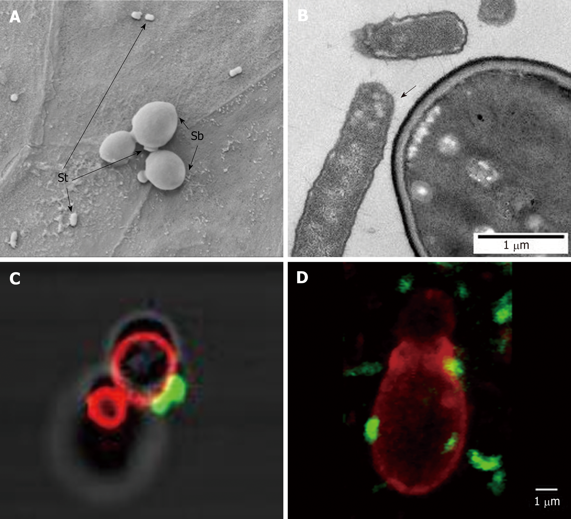
Figure 6 Scanning electron microscopy image showing Saccharomyces boulardii CNCM I-745 and Saccharomyces typhimurium on a monolayer of T84 polarized cells.
A and B: Electron microscopy image showing Saccharomyces typhimurium adhesion to Saccharomyces boulardii CNCM I-745; C and D: Confocal microscopy images showing S. typhimurium (Fluorescein IsoThioCyanate labelling), which adheres to S. boulardii CNCM I-745 (rhodamine labeling) in vitro (C) and in vivo on mouse cecum sections (D). Photos A and B: D. Czerucka1, P. Gounon2, P. Rampal1; C and D: D. Czerucka1, R. Pontier-Bres1, P. Rampal1 (1CSM, Monaco, microscopy Platform Cote d’Azur, MICA; 2University of Nice-Sophia-Antipolis). Sb: Saccharomyces boulardii CNCM I-745; St: Salmonella typhimurium.
- Citation: Czerucka D, Rampal P. Diversity of Saccharomyces boulardii CNCM I-745 mechanisms of action against intestinal infections. World J Gastroenterol 2019; 25(18): 2188-2203
- URL: https://www.wjgnet.com/1007-9327/full/v25/i18/2188.htm
- DOI: https://dx.doi.org/10.3748/wjg.v25.i18.2188


