Copyright
©The Author(s) 2019.
World J Gastroenterol. Apr 28, 2019; 25(16): 1997-2009
Published online Apr 28, 2019. doi: 10.3748/wjg.v25.i16.1997
Published online Apr 28, 2019. doi: 10.3748/wjg.v25.i16.1997
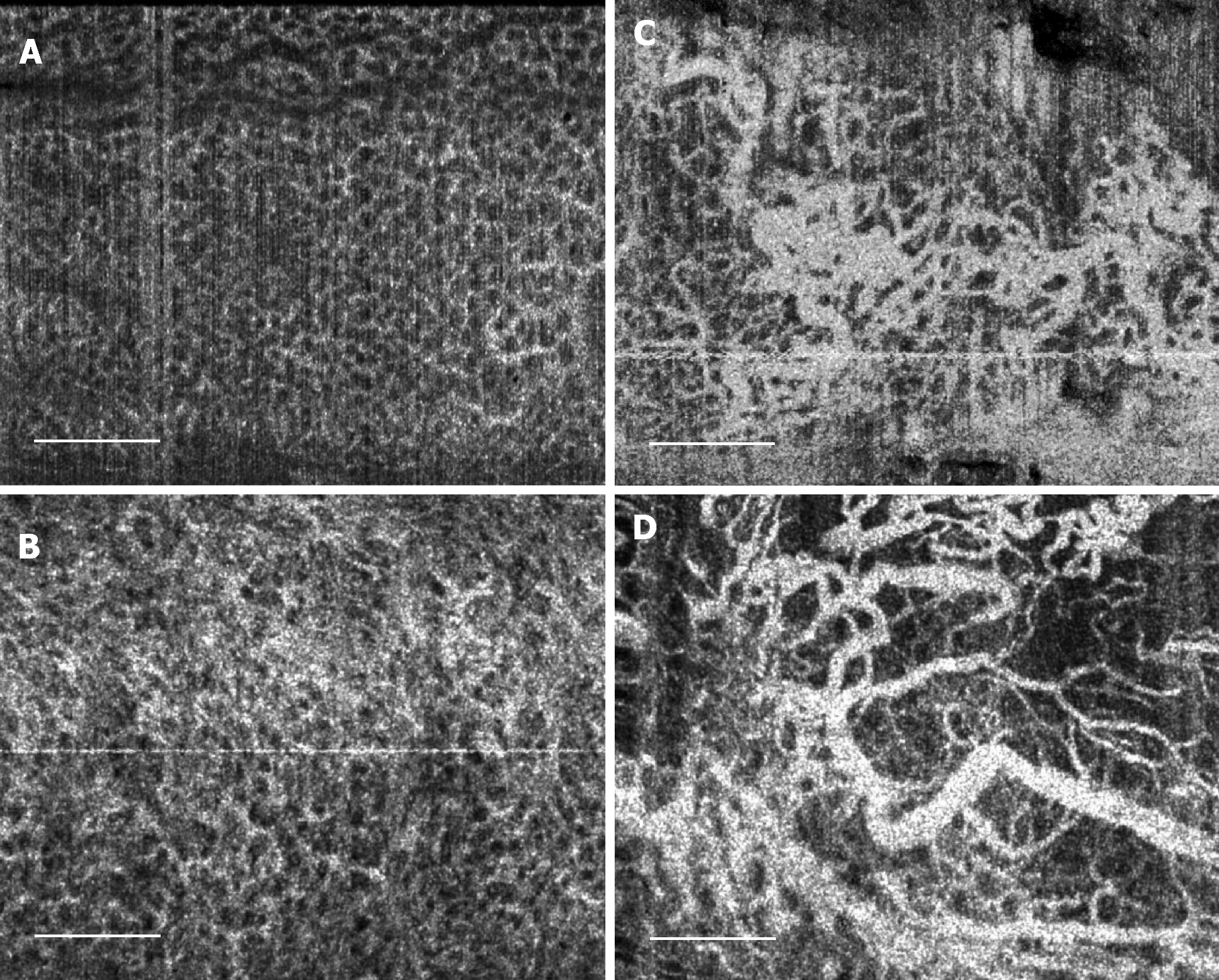
Figure 4 Summary of normal and abnormal microvasculature in the mucosal and submucosal layers of the rectum.
A: Normal rectal mucosal microvasculature consisted of a honeycomb-like microvascular pattern corresponding to subsurface capillary network; B: Abnormal rectal mucosal microvasculature had distortions to the honeycomb-like microvascular pattern, and had ectatic and tortuous microvasculature; C: Normal rectal submucosal microvasculature consisted of arterioles and venules with homogeneous vessel diameters typically of < 200 μm, in addition to shadowing from the superficial mucosal microvasculature; D: Abnormal rectal submucosal microvasculature had arterioles and venules with heterogonous and unusually high vessel diameters (> 200 μm). Scale bars are 1 mm.
- Citation: Ahsen OO, Liang K, Lee HC, Wang Z, Fujimoto JG, Mashimo H. Assessment of chronic radiation proctopathy and radiofrequency ablation treatment follow-up with optical coherence tomography angiography: A pilot study. World J Gastroenterol 2019; 25(16): 1997-2009
- URL: https://www.wjgnet.com/1007-9327/full/v25/i16/1997.htm
- DOI: https://dx.doi.org/10.3748/wjg.v25.i16.1997


