Copyright
©The Author(s) 2019.
World J Gastroenterol. Apr 28, 2019; 25(16): 1928-1935
Published online Apr 28, 2019. doi: 10.3748/wjg.v25.i16.1928
Published online Apr 28, 2019. doi: 10.3748/wjg.v25.i16.1928
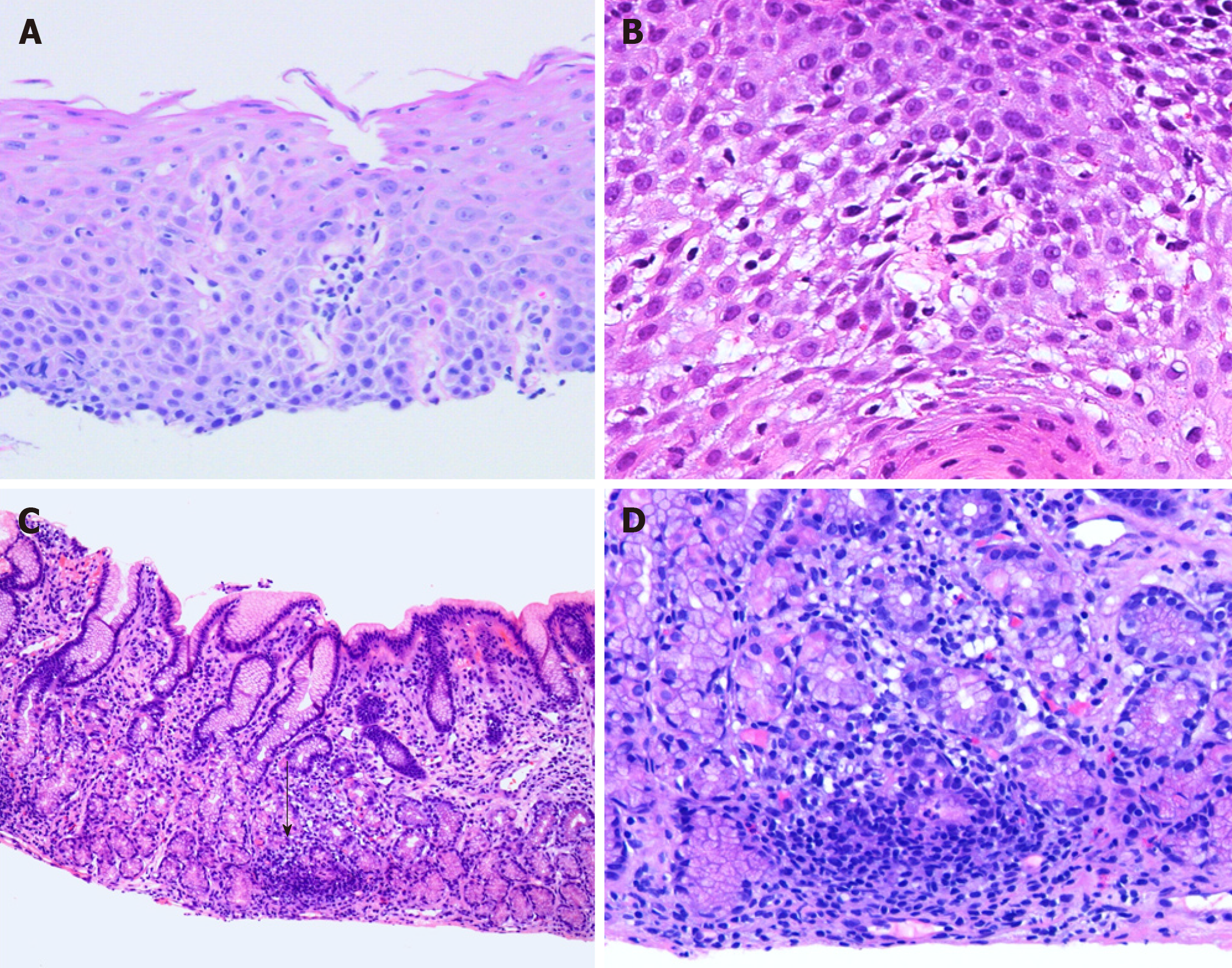
Figure 1 Hematoxylin and eosin staining.
A and B: Lymphocytic esophagitis is characterized by increased intraepithelial lymphocytes, predominantly peripapillary, with absence of intraepithelial eosinophils and neutrophils [hematoxylin and eosin (H and E), 10 × and 20 ×]. C: Low power view of focally enhanced gastritis demonstrates focal dense infiltrate surrounding a gland (black arrow) with a minimal increase of lymphoplasmacytic infiltrate in the lamina propria (H and E, 4 ×); D: Focally enhanced gastritis is characterized by lymphohistiocytic infiltrate with scattered neutrophils and associated glandular damage seen on medium power view (H and E, 10 ×).
- Citation: Abuquteish D, Putra J. Upper gastrointestinal tract involvement of pediatric inflammatory bowel disease: A pathological review. World J Gastroenterol 2019; 25(16): 1928-1935
- URL: https://www.wjgnet.com/1007-9327/full/v25/i16/1928.htm
- DOI: https://dx.doi.org/10.3748/wjg.v25.i16.1928


