Copyright
©The Author(s) 2019.
World J Gastroenterol. Apr 7, 2019; 25(13): 1531-1549
Published online Apr 7, 2019. doi: 10.3748/wjg.v25.i13.1531
Published online Apr 7, 2019. doi: 10.3748/wjg.v25.i13.1531
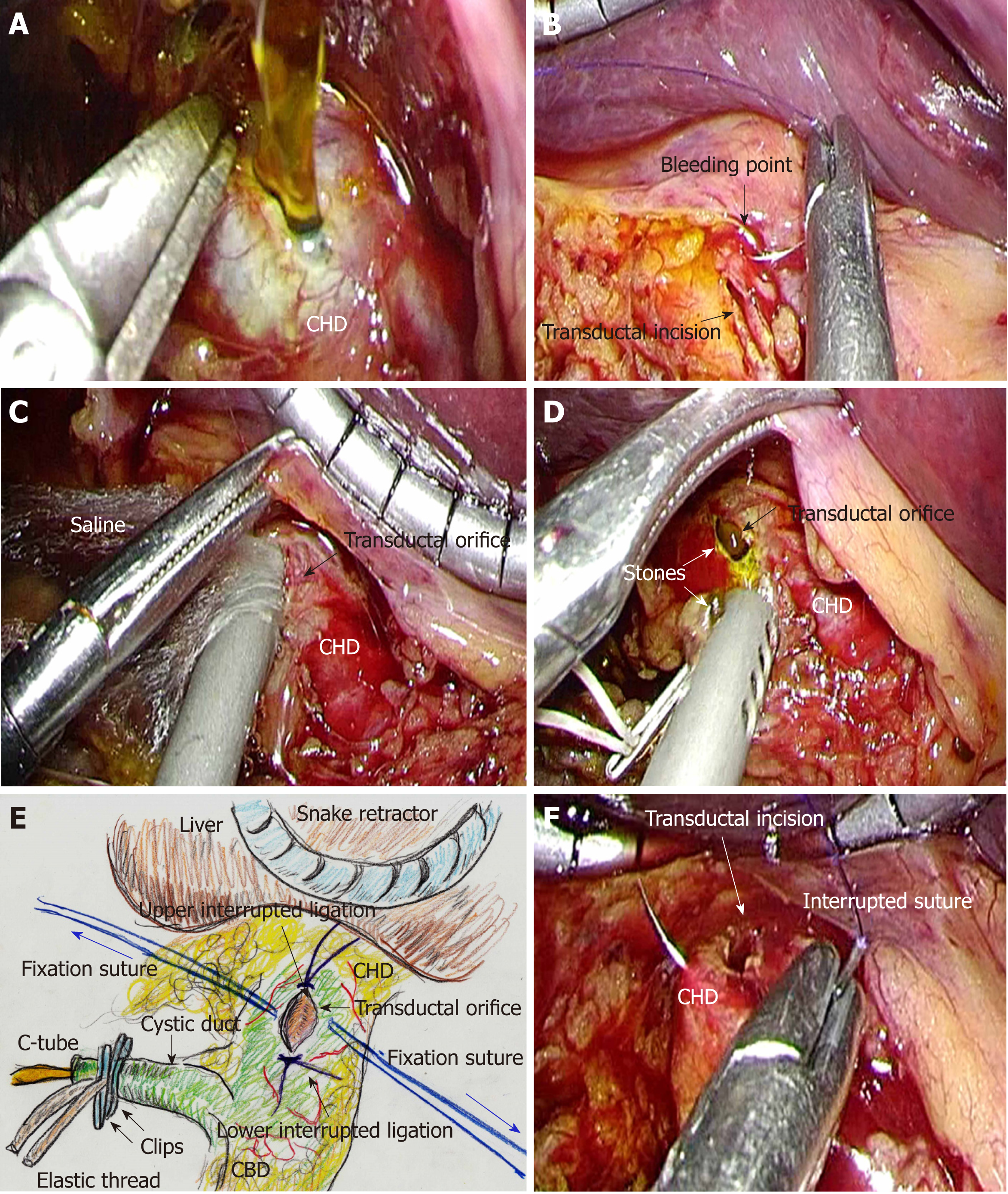
Figure 4 Actual surgical procedures of laparoscopic choledocholithotomy.
A: The extrahepatic bile duct (EHBD) is opened with sharp dissection; B: Intracorporeal suture placement and subsequent ligation are the first choice for hemostasis. No energy devices should be used; C: The cavity of the EHBD is sufficiently flushed. Frequent continuous suction is needed during laparoscopic choledocholithotomy. An automatically maintained pneumoperitoneum system is used to preserve an adequate surgical field; D: All stones are removed; E: Interrupted sutures are placed and subsequently ligated at the upper and lower edges of the transductal orifice to prevent progressive laceration resulting from cholangioscopic maneuvers. Thereafter, fixation sutures (blue arrows) are bilaterally placed to open the transductal orifice. These fixation sutures are adequately set through the abdominal wall at different points from the laparoscopic trocars; F: Interrupted sutures and subsequent ligation are placed at the upper and lower edges of the transductal incision, to prevent progressive laceration due to cholangioscopic maneuvers. CHD: Common hepatic duct; CBD: Common bile duct; EHBD: Extrahepatic bile duct.
- Citation: Hori T. Comprehensive and innovative techniques for laparoscopic choledocholithotomy: A surgical guide to successfully accomplish this advanced manipulation. World J Gastroenterol 2019; 25(13): 1531-1549
- URL: https://www.wjgnet.com/1007-9327/full/v25/i13/1531.htm
- DOI: https://dx.doi.org/10.3748/wjg.v25.i13.1531


