Copyright
©The Author(s) 2019.
World J Gastroenterol. Apr 7, 2019; 25(13): 1531-1549
Published online Apr 7, 2019. doi: 10.3748/wjg.v25.i13.1531
Published online Apr 7, 2019. doi: 10.3748/wjg.v25.i13.1531
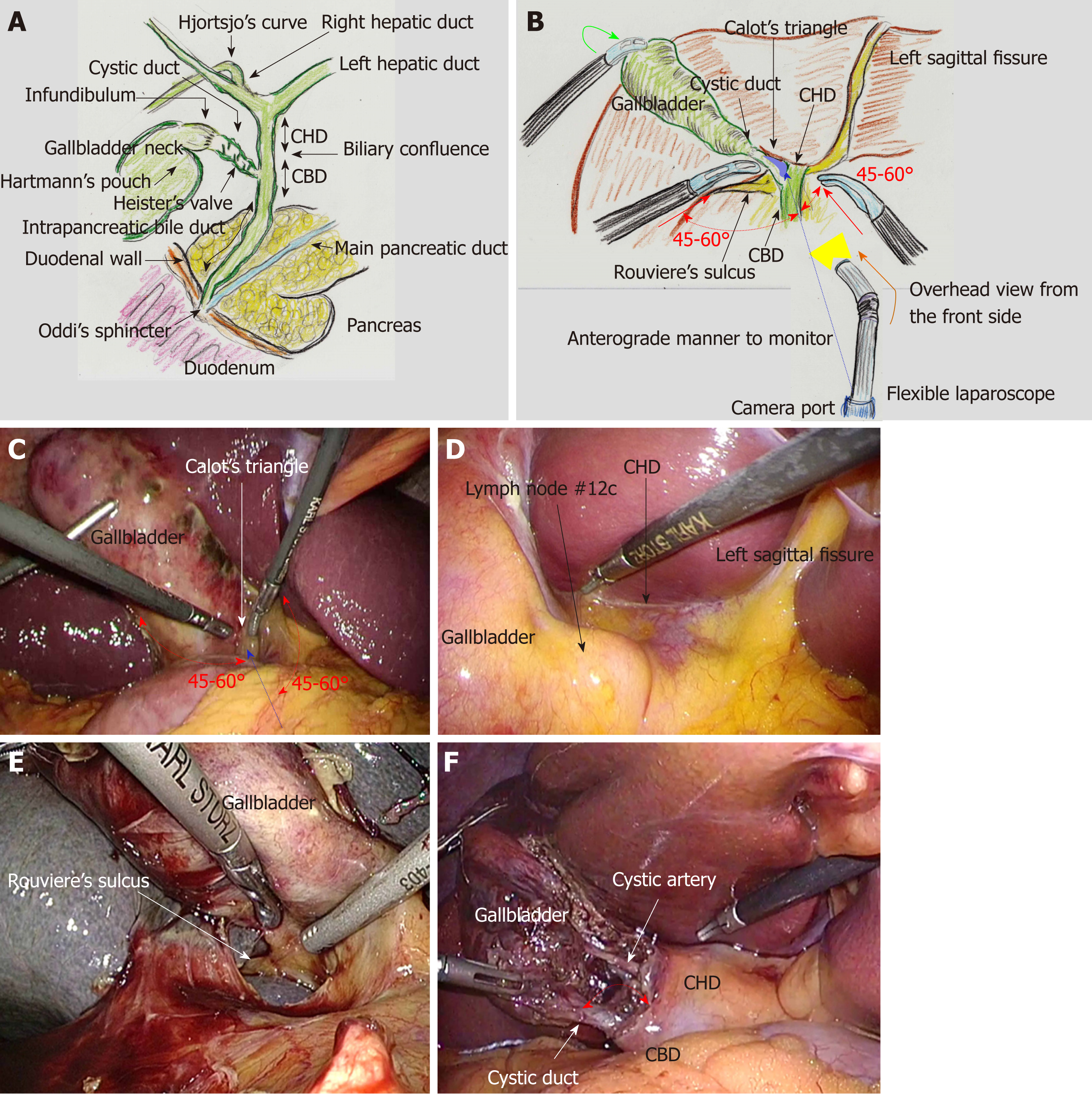
Figure 1 Biliary system and actual surgical procedures of laparoscopic choledocholithotomy.
A: The common hepatic duct (CHD), common bile duct (CBD) and intra-pancreatic bile duct compose the extrahepatic bile duct. Biliary drainage is regulated by Oddi’s sphincter. Recognition of Hjortsjo’s curve on cholangiography is useful for detecting the posterior branch from the right hepatic duct; B and C: The gallbladder fundus is superiorly and cranially lifted (green arrow). The target site is Calot’s triangle (blue shaded area). The two forceps of the main surgeon (red arrow) form appropriate angles (approximately 45°-60°) (red dotted arrow) to the axis from the camera port to Calot’s triangle (blue dotted arrow). A flexible laparoscope provides an overhead view from the upper anterior side (orange arrow), anterograde to the visual monitor; D: The bottom plateau of the U-shaped line from the left sagittal fissure to the gallbladder, which necessarily involves the CHD; E: Rouviere’s sulcus always involves the right hepatic duct; F: The whiter color change of the cystic duct is recognized, and a wider angle is created between the cystic duct and CHD (red arrow). CHD: Common hepatic duct; CBD: Common bile duct.
- Citation: Hori T. Comprehensive and innovative techniques for laparoscopic choledocholithotomy: A surgical guide to successfully accomplish this advanced manipulation. World J Gastroenterol 2019; 25(13): 1531-1549
- URL: https://www.wjgnet.com/1007-9327/full/v25/i13/1531.htm
- DOI: https://dx.doi.org/10.3748/wjg.v25.i13.1531


