Copyright
©The Author(s) 2018.
World J Gastroenterol. Feb 21, 2018; 24(7): 775-793
Published online Feb 21, 2018. doi: 10.3748/wjg.v24.i7.775
Published online Feb 21, 2018. doi: 10.3748/wjg.v24.i7.775
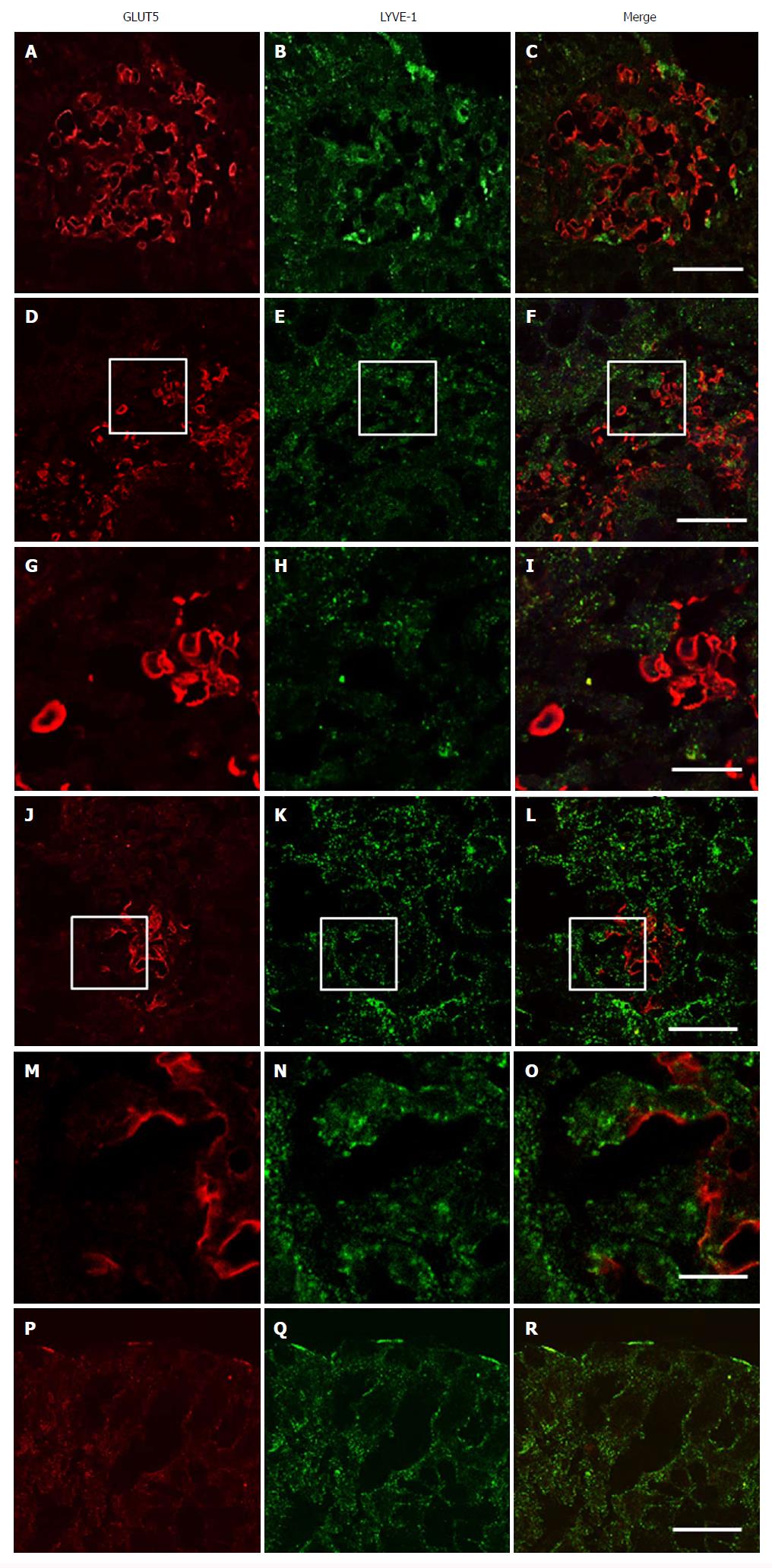
Figure 10 Double-immunofluorescent confocal microscopy showing expression of GLUT5 (red) with LYVE-1 (green) in samples of descending colon from UC (A-C, J-R) and CD (D-I) patients.
The mucosa was inflamed in the UC sample and non-inflamed in the CD sample. The boxed area in (D-F) is shown at higher magnification in (G-I, respectively); the boxed area in (J-L) is shown at higher magnification in (M-O, respectively). Scale bar: (C, F, L, R) 20 μm; (I, O) 10 μm.
- Citation: Merigo F, Brandolese A, Facchin S, Missaggia S, Bernardi P, Boschi F, D’Incà R, Savarino EV, Sbarbati A, Sturniolo GC. Glucose transporter expression in the human colon. World J Gastroenterol 2018; 24(7): 775-793
- URL: https://www.wjgnet.com/1007-9327/full/v24/i7/775.htm
- DOI: https://dx.doi.org/10.3748/wjg.v24.i7.775


