Copyright
©The Author(s) 2018.
World J Gastroenterol. Feb 21, 2018; 24(7): 775-793
Published online Feb 21, 2018. doi: 10.3748/wjg.v24.i7.775
Published online Feb 21, 2018. doi: 10.3748/wjg.v24.i7.775
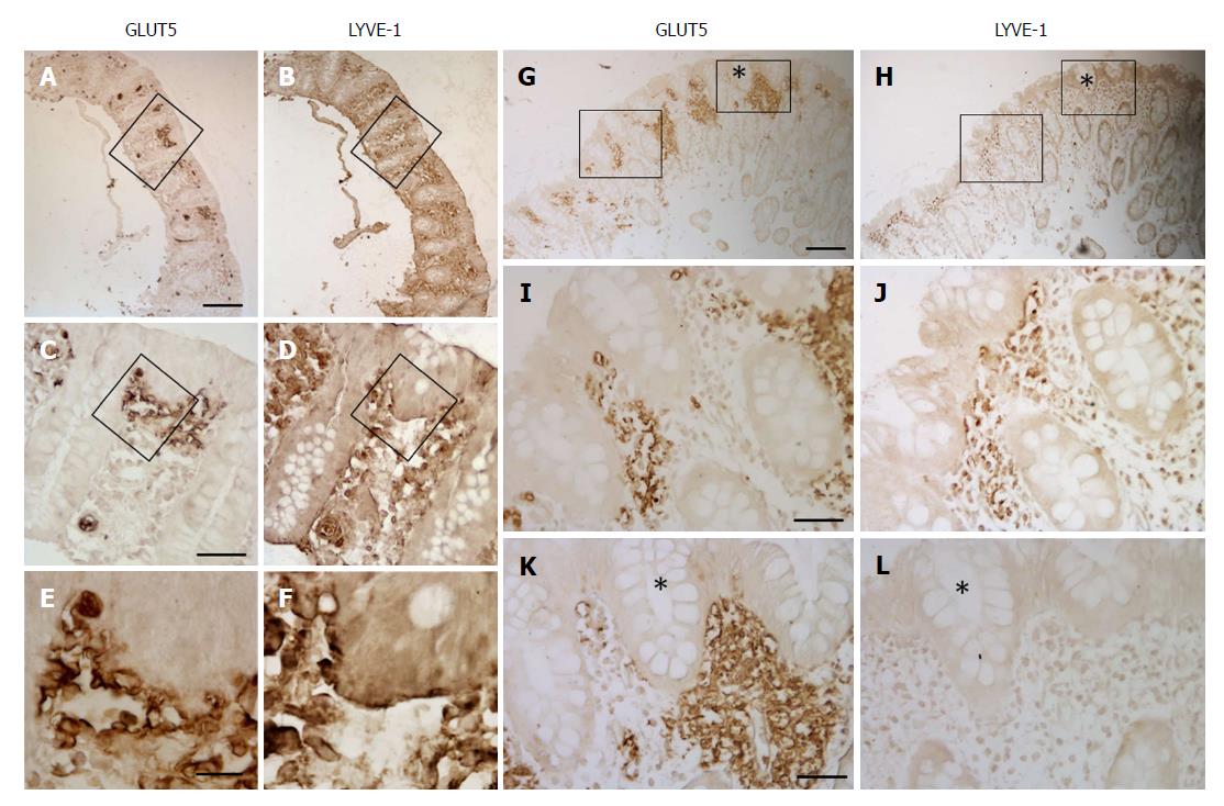
Figure 9 Immunoperoxidase staining showing GLUT5 and LYVE-1 immunoreactivity in samples of the distal tract of large intestine from UC (A-F) and CD (G-L) patients.
In both samples, the mucosa was non- inflamed. The boxed area in (A and B) is enlarged in (C and D, respectively), the area in (C and D) is enlarged in (E and F, respectively), the area without an asterisk in (G and H) is enlarged in (I and J, respectively), the area with an asterisk in (G and H) is enlarged in (K and L, respectively). Scale bar: (A, B, G, H) 100 μm; (C, D, I-L) 25 μm; (E and F) 10 μm.
- Citation: Merigo F, Brandolese A, Facchin S, Missaggia S, Bernardi P, Boschi F, D’Incà R, Savarino EV, Sbarbati A, Sturniolo GC. Glucose transporter expression in the human colon. World J Gastroenterol 2018; 24(7): 775-793
- URL: https://www.wjgnet.com/1007-9327/full/v24/i7/775.htm
- DOI: https://dx.doi.org/10.3748/wjg.v24.i7.775


