Copyright
©The Author(s) 2018.
World J Gastroenterol. Feb 7, 2018; 24(5): 651-656
Published online Feb 7, 2018. doi: 10.3748/wjg.v24.i5.651
Published online Feb 7, 2018. doi: 10.3748/wjg.v24.i5.651
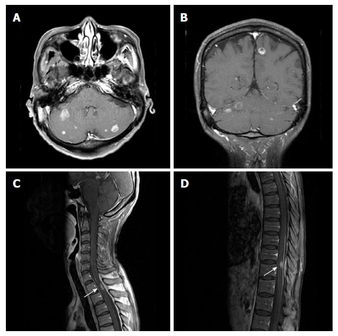
Figure 4 Magnetic resonance imaging of the brain and spine.
On axial (A) and coronal (B) contrast-enhanced T1-weighted imaging of the brain MRI revealed multiple enhancing nodules at the cerebral and cerebellar hemispheres. On sagittal contrast-enhanced T1-weighted imaging of the spine MRI revealed eccentric nodular enhancement at spinal cord at T1/2 (C) and T10/11 (D). MRI: Magnetic resonance imaging.
- Citation: Kim H, Yi KS, Kim WD, Son SM, Yang Y, Kwon J, Han HS. Sequential spinal and intracranial dural metastases in gastric adenocarcinoma: A case report. World J Gastroenterol 2018; 24(5): 651-656
- URL: https://www.wjgnet.com/1007-9327/full/v24/i5/651.htm
- DOI: https://dx.doi.org/10.3748/wjg.v24.i5.651


