Copyright
©The Author(s) 2018.
World J Gastroenterol. Dec 21, 2018; 24(47): 5379-5390
Published online Dec 21, 2018. doi: 10.3748/wjg.v24.i47.5379
Published online Dec 21, 2018. doi: 10.3748/wjg.v24.i47.5379
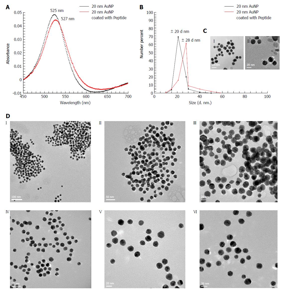
Figure 2 Characterization of peptide coated AuNPs.
A: Characterization of AuNP coated with peptide using a UV-Vis spectrophotometer indicating a spectral red shift in wavelength from 525 nm (for 20 nm AuNP only) to 527 nm (for 20 nm AuNP coated with peptide); B: Characterization of AuNP coated with peptide using DLS that showed an increase in the hydrodynamic size of the uncoated vs coated particles from 20 nm to 28 nm respectively; C: High resolution TEM images of (1) uncoated AuNPs, (2) AuNPs coated with peptide showing a “halo” layer surrounding the surface of the nanoparticles indicating coating of the gold with the peptide had occurred. In contrast, the “halo” effect was not observed on the surface of the un-coated AuNPs; D: High resolution TEM images following the incubation with AGA (12 μg/mL) (1, 2, 3) AuNPs coated with peptide showing aggregation confirming coating of peptide on AuNP (4, 5, 6) Uncoated AuNPs remained dispersed. DLS: Dynamic light scattering; TEM: Transmission electron microscopy.
- Citation: Kaur A, Shimoni O, Wallach M. Novel screening test for celiac disease using peptide functionalised gold nanoparticles. World J Gastroenterol 2018; 24(47): 5379-5390
- URL: https://www.wjgnet.com/1007-9327/full/v24/i47/5379.htm
- DOI: https://dx.doi.org/10.3748/wjg.v24.i47.5379


