Copyright
©The Author(s) 2018.
World J Gastroenterol. Dec 14, 2018; 24(46): 5215-5222
Published online Dec 14, 2018. doi: 10.3748/wjg.v24.i46.5215
Published online Dec 14, 2018. doi: 10.3748/wjg.v24.i46.5215
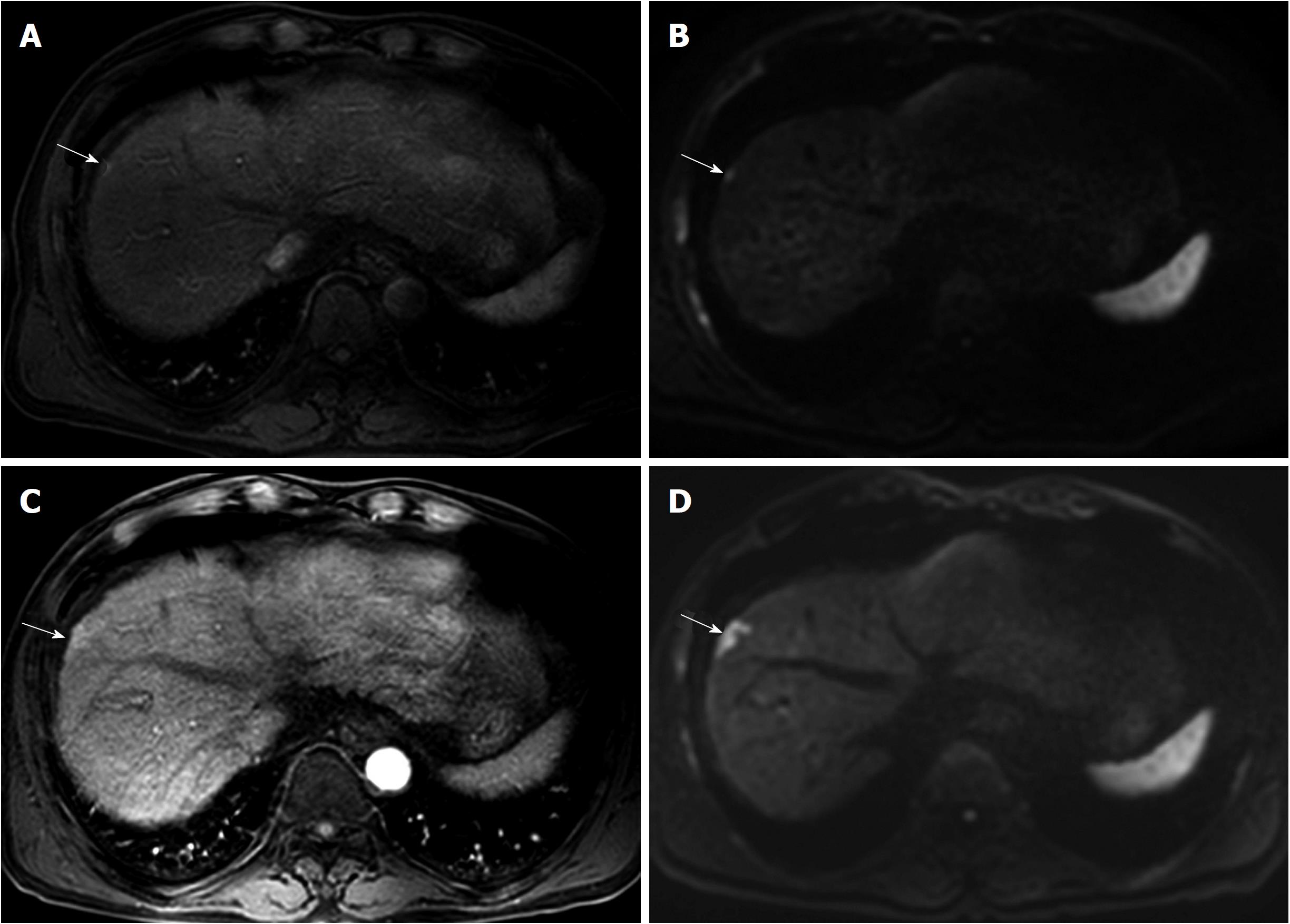
Figure 3 61-year-old man with a history of transarterial chemoembolization for hepatocellular carcinoma.
A 6-mm recurrent hepatocellular carcinoma in segment 8 of the liver is seen by magnetic resonance imaging (MRI). The tiny nodule (arrow) shows arterial enhancement (A) and high signal intensity on diffusion-weighted image (B). Unfortunately, there was no mention of the lesion in the radiologic report, and the tumor was unintentionally diagnosed late. At follow up MRI obtained 6 mo later, the lesion (arrow) had increased in size and measured up to 22 mm on arterial phase (C) and diffusion-weighted image (D). Given that the lesion had irregular margins and showed rapid growth, the tumor might have aggressive tumor biology.
- Citation: Lee MW, Lim HK. Management of sub-centimeter recurrent hepatocellular carcinoma after curative treatment: Current status and future. World J Gastroenterol 2018; 24(46): 5215-5222
- URL: https://www.wjgnet.com/1007-9327/full/v24/i46/5215.htm
- DOI: https://dx.doi.org/10.3748/wjg.v24.i46.5215


