Copyright
©The Author(s) 2018.
World J Gastroenterol. Dec 14, 2018; 24(46): 5215-5222
Published online Dec 14, 2018. doi: 10.3748/wjg.v24.i46.5215
Published online Dec 14, 2018. doi: 10.3748/wjg.v24.i46.5215
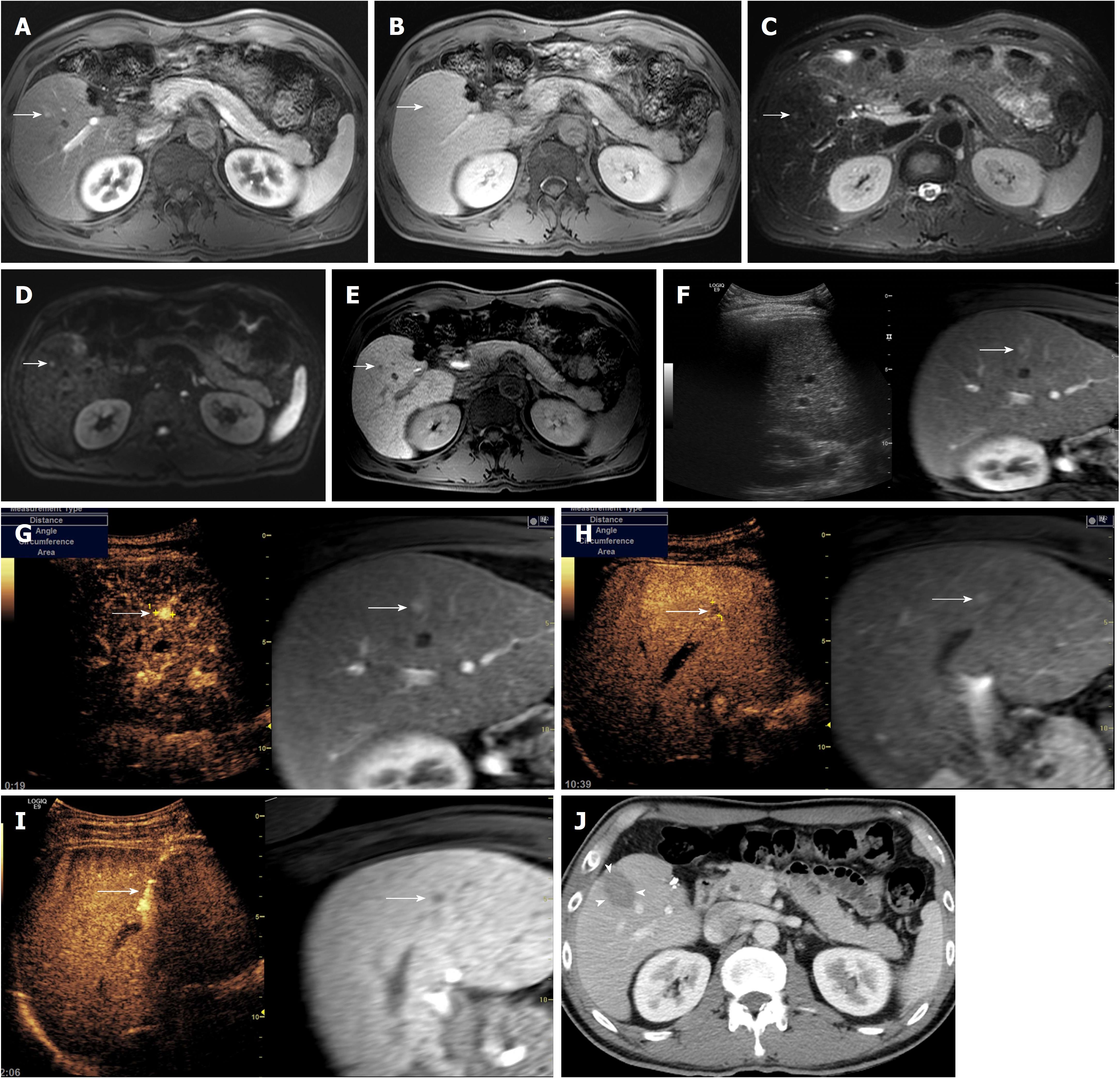
Figure 1 45-year-old man with a history of left hemihepatectomy for hepatocellular carcinoma.
A 6-mm recurrent hepatocellular carcinoma (HCC) in segment 5 of the liver is seen on magnetic resonance imaging. The nodule (arrow) shows arterial enhancement (A), washout on transitional phase (B), high signal intensity on both T2- and diffusion-weighted images (C and D), and low signal intensity on hepatobiliary phase (E). On planning ultrasound (US) with fusion imaging (F), the nodule was invisible using real-time US image (left figure) at the corresponding site of the fused magnetic resonance image (right figure). When contrast-enhanced ultrasound (CEUS) using Sonazoid (GE Healthcare, Oslo, Norway) was added to fusion imaging, there was a nodule (arrow) with arterial enhancement (G) and low echogenicity in the post-vascular phase (H). Under CEUS-added fusion imaging guidance (I), the radiofrequency (RF) electrode was accurately placed through the tumor (arrow). At follow up computed tomography obtained immediately after RF ablation (J), the index tumor was completely ablated with sufficient ablative margin (arrowhead).
- Citation: Lee MW, Lim HK. Management of sub-centimeter recurrent hepatocellular carcinoma after curative treatment: Current status and future. World J Gastroenterol 2018; 24(46): 5215-5222
- URL: https://www.wjgnet.com/1007-9327/full/v24/i46/5215.htm
- DOI: https://dx.doi.org/10.3748/wjg.v24.i46.5215


