Copyright
©The Author(s) 2018.
World J Gastroenterol. Dec 7, 2018; 24(45): 5109-5119
Published online Dec 7, 2018. doi: 10.3748/wjg.v24.i45.5109
Published online Dec 7, 2018. doi: 10.3748/wjg.v24.i45.5109
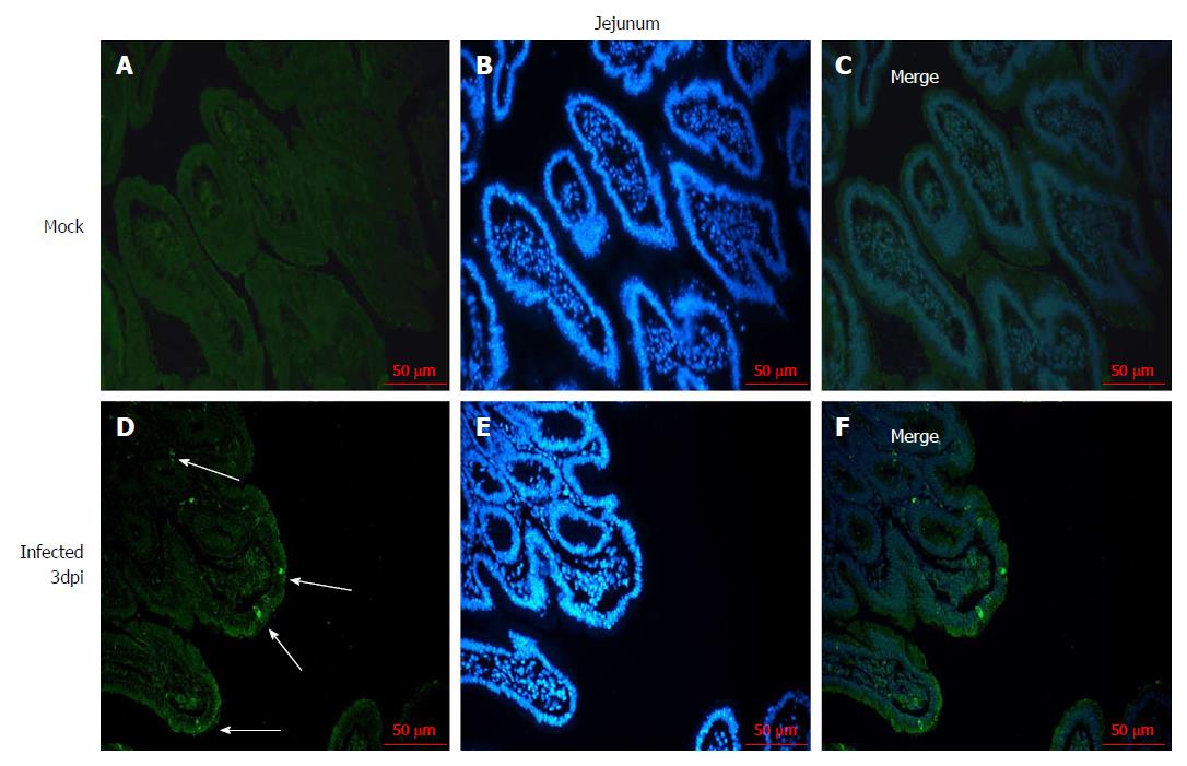
Figure 2 Immunofluorescence of rotavirus antigen in the jejunum of neonatal rhesus monkeys inoculated with SA11 or medium without serum.
A-C: The jejunum of neonatal rhesus monkeys inoculated with medium without serum at 3 dpi; D-F: Jejunum of neonatal rhesus monkeys inoculated with 108 PFUs of SA11/monkey at 3 dpi. The glass slides were incubated with goat anti-rotavirus (RV) polyclonal antibody and then incubated with rabbit anti-goat IgG antibody labeled with FITC (green). Cell nuclei are shown with DAPI staining (blue). White arrows indicate representative RV-positive cells. Magnification, × 20. Bar: 50 μm.
- Citation: Yin N, Yang FM, Qiao HT, Zhou Y, Duan SQ, Lin XC, Wu JY, Xie YP, He ZL, Sun MS, Li HJ. Neonatal rhesus monkeys as an animal model for rotavirus infection. World J Gastroenterol 2018; 24(45): 5109-5119
- URL: https://www.wjgnet.com/1007-9327/full/v24/i45/5109.htm
- DOI: https://dx.doi.org/10.3748/wjg.v24.i45.5109


