Copyright
©The Author(s) 2018.
World J Gastroenterol. Nov 28, 2018; 24(44): 5005-5012
Published online Nov 28, 2018. doi: 10.3748/wjg.v24.i44.5005
Published online Nov 28, 2018. doi: 10.3748/wjg.v24.i44.5005
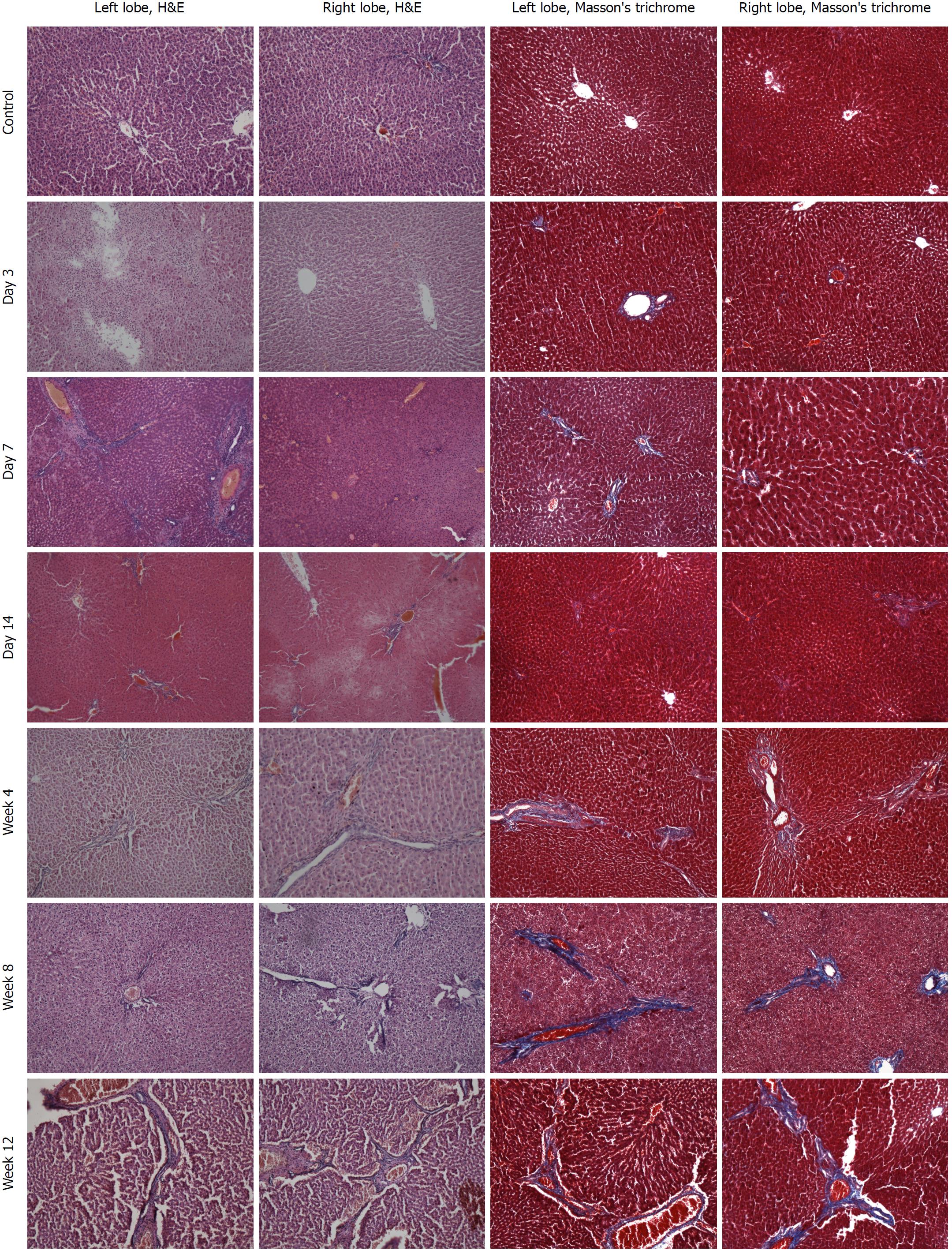
Figure 2 Histopathological changes of the liver (× 100).
H&E staining of sections of the left lobe (1st column) and right lobe (2nd column) of the liver, and Masson’s trichrome staining of sections of the left lobe (3rd column) and right lobe (4th column) of the liver were performed in the control group (1st row) and at each time point after injection of colchicine (2nd to 7th rows). No obvious pathological changes were observed in the control group. The hepatic injury was different between the left and right lobes at each time point. At day 3 after colchicine injection, there was massive inflammatory cells infiltration, hepatocellular edema, and mild liver necrosis. At day 14, reduced inflammation and increased necrosis were observed, while fibrosis was not detected. At week 4, cholestasis and early fibrosis were observed. At weeks 8 and 12, there was further fibrosis.
- Citation: Zhang YY, Zhang CX, Li Y, Jiang X, Wang YF, Sun Y, Wang J, Ji WY, Liu Y. Development of a novel rat model of heterogeneous hepatic injury by injection with colchicine via the splenic vein. World J Gastroenterol 2018; 24(44): 5005-5012
- URL: https://www.wjgnet.com/1007-9327/full/v24/i44/5005.htm
- DOI: https://dx.doi.org/10.3748/wjg.v24.i44.5005


