Copyright
©The Author(s) 2018.
World J Gastroenterol. Nov 21, 2018; 24(43): 4880-4892
Published online Nov 21, 2018. doi: 10.3748/wjg.v24.i43.4880
Published online Nov 21, 2018. doi: 10.3748/wjg.v24.i43.4880
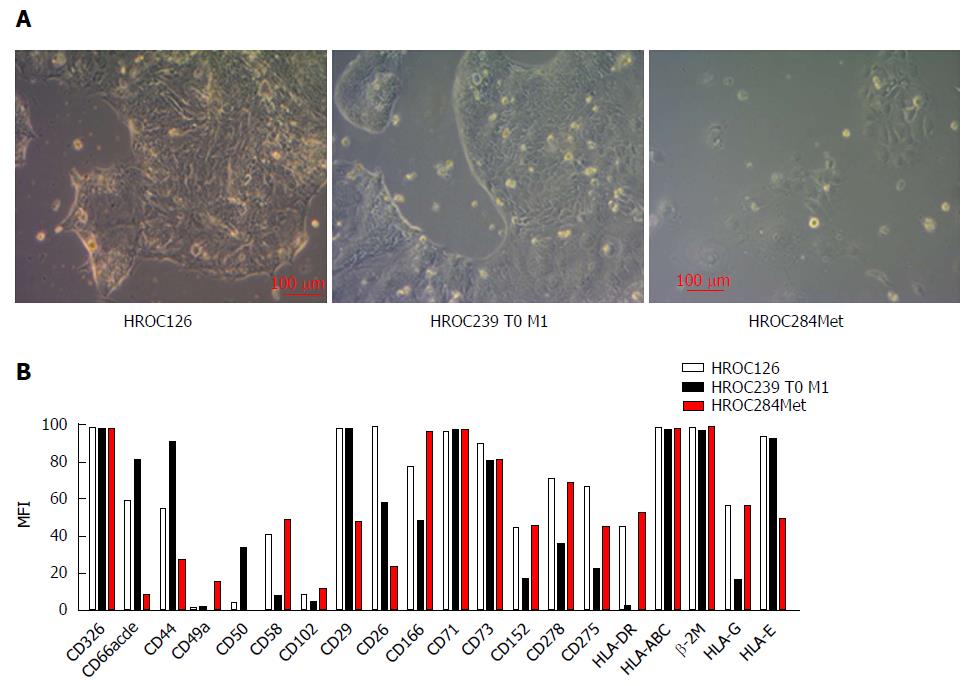
Figure 2 Morphology and phenotype of individual rectal cancer cell lines.
A: Light microscopy of freshly established tumor cell lines (all passage 6-11). Cell lines were established from patients’ tumor material as described in material and methods. Original magnification × 100; B: Phenotyping was conducted by flow cytometry (BD FACSARIA II) using fluorochrome-labeled mAbs as given on the x-axis. Exemplary data of one analysis out of three biological replicates are given. Some markers display a high variation in dependence from cell density in the culture vessels. HLA: Human leukocyte antigen.
- Citation: Gock M, Mullins CS, Bergner C, Prall F, Ramer R, Göder A, Krämer OH, Lange F, Krause BJ, Klar E, Linnebacher M. Establishment, functional and genetic characterization of three novel patient-derived rectal cancer cell lines. World J Gastroenterol 2018; 24(43): 4880-4892
- URL: https://www.wjgnet.com/1007-9327/full/v24/i43/4880.htm
- DOI: https://dx.doi.org/10.3748/wjg.v24.i43.4880


