Copyright
©The Author(s) 2018.
World J Gastroenterol. Nov 14, 2018; 24(42): 4809-4820
Published online Nov 14, 2018. doi: 10.3748/wjg.v24.i42.4809
Published online Nov 14, 2018. doi: 10.3748/wjg.v24.i42.4809
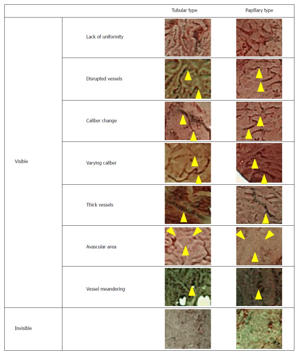
Figure 3 Vascular patterns of colorectal tumors as revealed by narrow-band imaging endoscopy.
Colorectal lesions were broadly classified into tubular or papillary types based on narrow-band imaging observations of the histopathologic appearances and proliferation patterns, followed by an evaluation of the microvessel architecture. “Disturbed arrangement” was defined as a disturbance in the form, size, and arrangement of microvessels; “disrupted vessels” was defined as an evident disruption of the microvessels; “varied caliber” was defined as a variance of caliber in the same vessel; “size irregularity” was defined as variance in caliber among different vessels; “thick vessels” was defined as an area harboring cyan-colored vessels relative to vessels in adjacent areas; “avascular area” was defined as an area with no visible vessels; and “vessel meandering” was defined as evident meandering of the blood vessels along a twisting and winding path. Representative images are shown. The locations of vessels representative of each vascular pattern and classification are denoted by arrowheads. Magnification: 125 ×.
- Citation: Maeyama Y, Mitsuyama K, Noda T, Nagata S, Nagata T, Yoshioka S, Yoshida H, Mukasa M, Sumie H, Kawano H, Akiba J, Araki Y, Kakuma T, Tsuruta O, Torimura T. Prediction of colorectal tumor grade and invasion depth through narrow-band imaging scoring. World J Gastroenterol 2018; 24(42): 4809-4820
- URL: https://www.wjgnet.com/1007-9327/full/v24/i42/4809.htm
- DOI: https://dx.doi.org/10.3748/wjg.v24.i42.4809


