Copyright
©The Author(s) 2018.
World J Gastroenterol. Nov 14, 2018; 24(42): 4809-4820
Published online Nov 14, 2018. doi: 10.3748/wjg.v24.i42.4809
Published online Nov 14, 2018. doi: 10.3748/wjg.v24.i42.4809
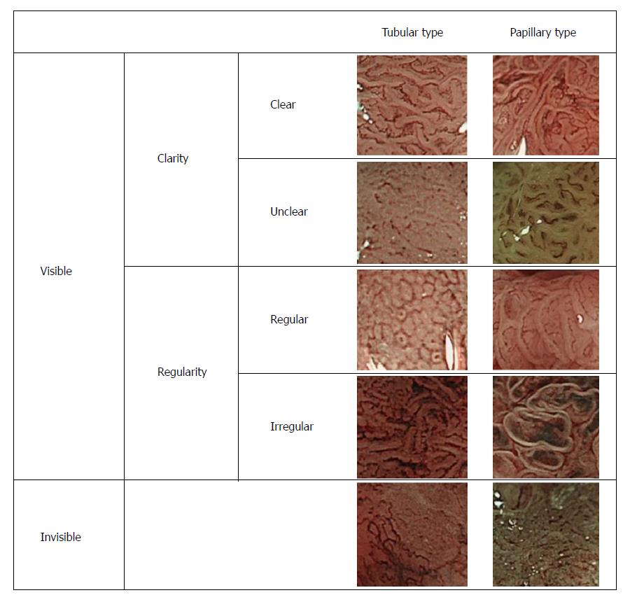
Figure 2 Classification of surface patterns of colorectal tumors as revealed by narrow-band imaging.
Colorectal lesions were broadly classified into tubular or papillary types, based on narrow-band imaging observations of histopathologic appearances and proliferation patterns; this was followed by an evaluation of the microstructure of the superficial layer of each lesion. Regarding the surface pattern, lesions with a clearly visualized surface microstructure were defined as the “clear type,” whereas lesions with a visualized but not readily discerned surface microstructure were defined as the “unclear type.” Lesions with a surface microstructure characterized by uniformly-sized pits in a regularly arranged pattern were defined as the “regular type,” whereas those with an irregular pit size and arrangement were defined as the “irregular type.” Representative images are shown. Magnification: 125 ×.
- Citation: Maeyama Y, Mitsuyama K, Noda T, Nagata S, Nagata T, Yoshioka S, Yoshida H, Mukasa M, Sumie H, Kawano H, Akiba J, Araki Y, Kakuma T, Tsuruta O, Torimura T. Prediction of colorectal tumor grade and invasion depth through narrow-band imaging scoring. World J Gastroenterol 2018; 24(42): 4809-4820
- URL: https://www.wjgnet.com/1007-9327/full/v24/i42/4809.htm
- DOI: https://dx.doi.org/10.3748/wjg.v24.i42.4809


