Copyright
©The Author(s) 2018.
World J Gastroenterol. Nov 14, 2018; 24(42): 4759-4772
Published online Nov 14, 2018. doi: 10.3748/wjg.v24.i42.4759
Published online Nov 14, 2018. doi: 10.3748/wjg.v24.i42.4759
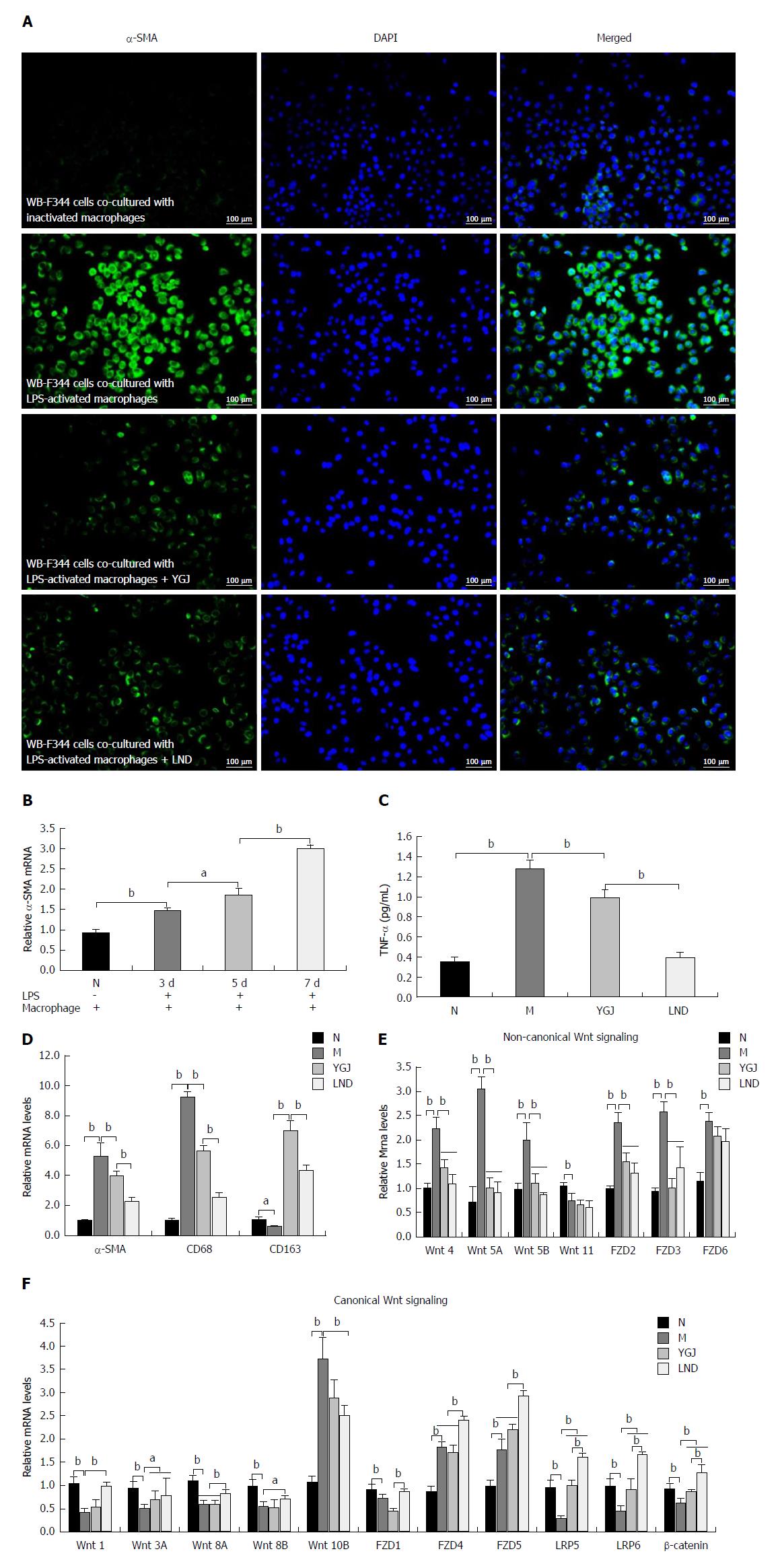
Figure 5 Activation of macrophages and differentiation of WB-F344 to myofibroblasts in vitro.
A: Double immunofluorescent staining of α-smooth muscle actin (α-SMA) (red) and DAPI (blue) merged (× 200). B: The mRNA expression of α-SMA in WB-F344 cells after co-culture with lipopolysaccharide (LPS)-activated RAW264.7 cells for 3, 5, or 7 d. C: Tumor necrosis factor production in LPS-activated RAW264.7 was detected by enzyme-linked immuno sorbent assay. D: Relative mRNA expression levels of α-SMA (WB-F344), CD68 (RAW264.7), and CD163 (RAW264.7). E: Relative mRNA levels of non-canonical Wnt signaling pathway components. F: Relative mRNA levels of canonical Wnt signaling pathway components. The mRNA levels were normalized to the GAPDH expression levels. aP < 0.05, bP < 0.01. N: Normal control group; M: WB-F344 cells co-cultured with LPS-activated RAW264.7 cells group; YGJ: Yiguanjian decoction group; LND: Lenalidomide group.
- Citation: Xu Y, Fan WW, Xu W, Jiang SL, Chen GF, Liu C, Chen JM, Zhang H, Liu P, Mu YP. Yiguanjian decoction enhances fetal liver stem/progenitor cell-mediated repair of liver cirrhosis through regulation of macrophage activation state. World J Gastroenterol 2018; 24(42): 4759-4772
- URL: https://www.wjgnet.com/1007-9327/full/v24/i42/4759.htm
- DOI: https://dx.doi.org/10.3748/wjg.v24.i42.4759


