Copyright
©The Author(s) 2018.
World J Gastroenterol. Jan 28, 2018; 24(4): 543-548
Published online Jan 28, 2018. doi: 10.3748/wjg.v24.i4.543
Published online Jan 28, 2018. doi: 10.3748/wjg.v24.i4.543
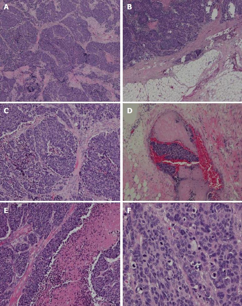
Figure 2 Histological findings.
A: A low-power histological view. Tumor cells show infiltrative growth from the muscularis propria to the subserosa (HE, × 40). B: Large-cell carcinoma showing invasion into the subserosa. C: High-power view shows monotonous large tumor cells with abundant cytoplasm and large irregular nuclei with prominent nucleoli (HE, × 100). D and E: Angiolymphatic invasion and carcinoma cell embolus. F: Mitotic figures were also observed (60 per 10 high-power fields). HE: Hematoxylin and eosin.
- Citation: Ma FH, Xue LY, Chen YT, Xie YB, Zhong YX, Xu Q, Tian YT. Neuroendocrine carcinoma of the gastric stump: A case report and literature review. World J Gastroenterol 2018; 24(4): 543-548
- URL: https://www.wjgnet.com/1007-9327/full/v24/i4/543.htm
- DOI: https://dx.doi.org/10.3748/wjg.v24.i4.543


