Copyright
©The Author(s) 2018.
World J Gastroenterol. Jan 28, 2018; 24(4): 537-542
Published online Jan 28, 2018. doi: 10.3748/wjg.v24.i4.537
Published online Jan 28, 2018. doi: 10.3748/wjg.v24.i4.537
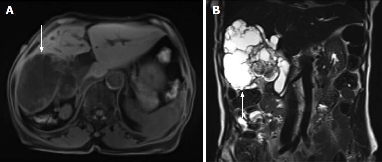
Figure 3 Magnetic resonance cholangiopancreatography.
A: A lobulated cystic mass in the right hepatic lobe with multifocal intramural enhancing nodules (white arrow); B: The connection of the cystic mass with the intrahepatic duct indicated a malignant transformation (white arrow). 300 mm × 225 mm (300 × 300 DPI).
- Citation: Lee JM, Lee JM, Hyun JJ, Choi HS, Kim ES, Keum B, Jeen YT, Chun HJ, Lee HS, Kim CD, Kim DS, Kim JY. Intraductal papillary bile duct adenocarcinoma and gastrointestinal stromal tumor in a case of neurofibromatosis type 1. World J Gastroenterol 2018; 24(4): 537-542
- URL: https://www.wjgnet.com/1007-9327/full/v24/i4/537.htm
- DOI: https://dx.doi.org/10.3748/wjg.v24.i4.537


