Copyright
©The Author(s) 2018.
World J Gastroenterol. Jan 28, 2018; 24(4): 461-474
Published online Jan 28, 2018. doi: 10.3748/wjg.v24.i4.461
Published online Jan 28, 2018. doi: 10.3748/wjg.v24.i4.461
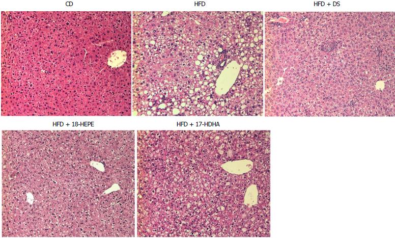
Figure 6 Liver tissue morphology.
Representative photomicrographs (Magnification × 20) Hematoxylin-eosin stain. HFD + DS showed a drastic decrease of number in fat vesicles and ballooning degeneration, but minimal change in inflammatory cells. Both groups HFD + 18-HEPE and HFD + 17-HDHA showed noticeable changes in steatosis and hepatocyte ballooning compared to HFD group. Plus, both groups displayed scarce presence of inflammatory cells.
- Citation: Rodriguez-Echevarria R, Macias-Barragan J, Parra-Vargas M, Davila-Rodriguez JR, Amezcua-Galvez E, Armendariz-Borunda J. Diet switch and omega-3 hydroxy-fatty acids display differential hepatoprotective effects in an obesity/nonalcoholic fatty liver disease model in mice. World J Gastroenterol 2018; 24(4): 461-474
- URL: https://www.wjgnet.com/1007-9327/full/v24/i4/461.htm
- DOI: https://dx.doi.org/10.3748/wjg.v24.i4.461


