Copyright
©The Author(s) 2018.
World J Gastroenterol. Oct 14, 2018; 24(38): 4341-4355
Published online Oct 14, 2018. doi: 10.3748/wjg.v24.i38.4341
Published online Oct 14, 2018. doi: 10.3748/wjg.v24.i38.4341
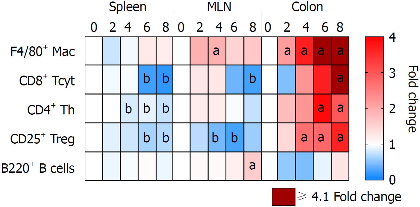
Figure 6 Immune cell population in the spleen, mesenteric lymph nodes and colon of 3% dextran sulfate sodium animals.
Tissue samples were collected from 3% DSS animals at different time points and analyzed through either immunohistochemistry or flow cytometry. The heatmap shows a possible movement of different immune cell types from the spleen and MLN into the gut progressively during the 8 d of disease. aP < 0.05 increased fold changes compared to control (day 0). bP < 0.05 decreased fold changes compared to control (day 0). MLN: Mesenteric lymph nodes; DSS: Dextran sulfate sodium.
- Citation: Nunes NS, Kim S, Sundby M, Chandran P, Burks SR, Paz AH, Frank JA. Temporal clinical, proteomic, histological and cellular immune responses of dextran sulfate sodium-induced acute colitis. World J Gastroenterol 2018; 24(38): 4341-4355
- URL: https://www.wjgnet.com/1007-9327/full/v24/i38/4341.htm
- DOI: https://dx.doi.org/10.3748/wjg.v24.i38.4341


