Copyright
©The Author(s) 2018.
World J Gastroenterol. Oct 7, 2018; 24(37): 4263-4271
Published online Oct 7, 2018. doi: 10.3748/wjg.v24.i37.4263
Published online Oct 7, 2018. doi: 10.3748/wjg.v24.i37.4263
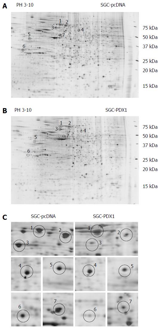
Figure 2 Two-dimensional electrophoresis-gel images of proteins from SGC7901 with pcDNA3.
1(+)-pancreatic-duodenal homeobox-1 (PDX1) vector and SGC7901-pcDNA cells. A and B: Representative 2DE-gel images of SGC-PDX1 and SGC-pcDNA groups. The differentially expressed proteins are indicated by circles; C: Close-up images of the differential protein spots between SGC-PDX1 and SGC-pcDNA groups. 2DE: 2-dimensional electrophoresis.
- Citation: Ma J, Wang BB, Ma XY, Deng WP, Xu LS, Sha WH. Potential involvement of heat shock proteins in pancreatic-duodenal homeobox-1-mediated effects on the genesis of gastric cancer: A 2D gel-based proteomic study. World J Gastroenterol 2018; 24(37): 4263-4271
- URL: https://www.wjgnet.com/1007-9327/full/v24/i37/4263.htm
- DOI: https://dx.doi.org/10.3748/wjg.v24.i37.4263


