Copyright
©The Author(s) 2018.
World J Gastroenterol. Sep 14, 2018; 24(34): 3861-3870
Published online Sep 14, 2018. doi: 10.3748/wjg.v24.i34.3861
Published online Sep 14, 2018. doi: 10.3748/wjg.v24.i34.3861
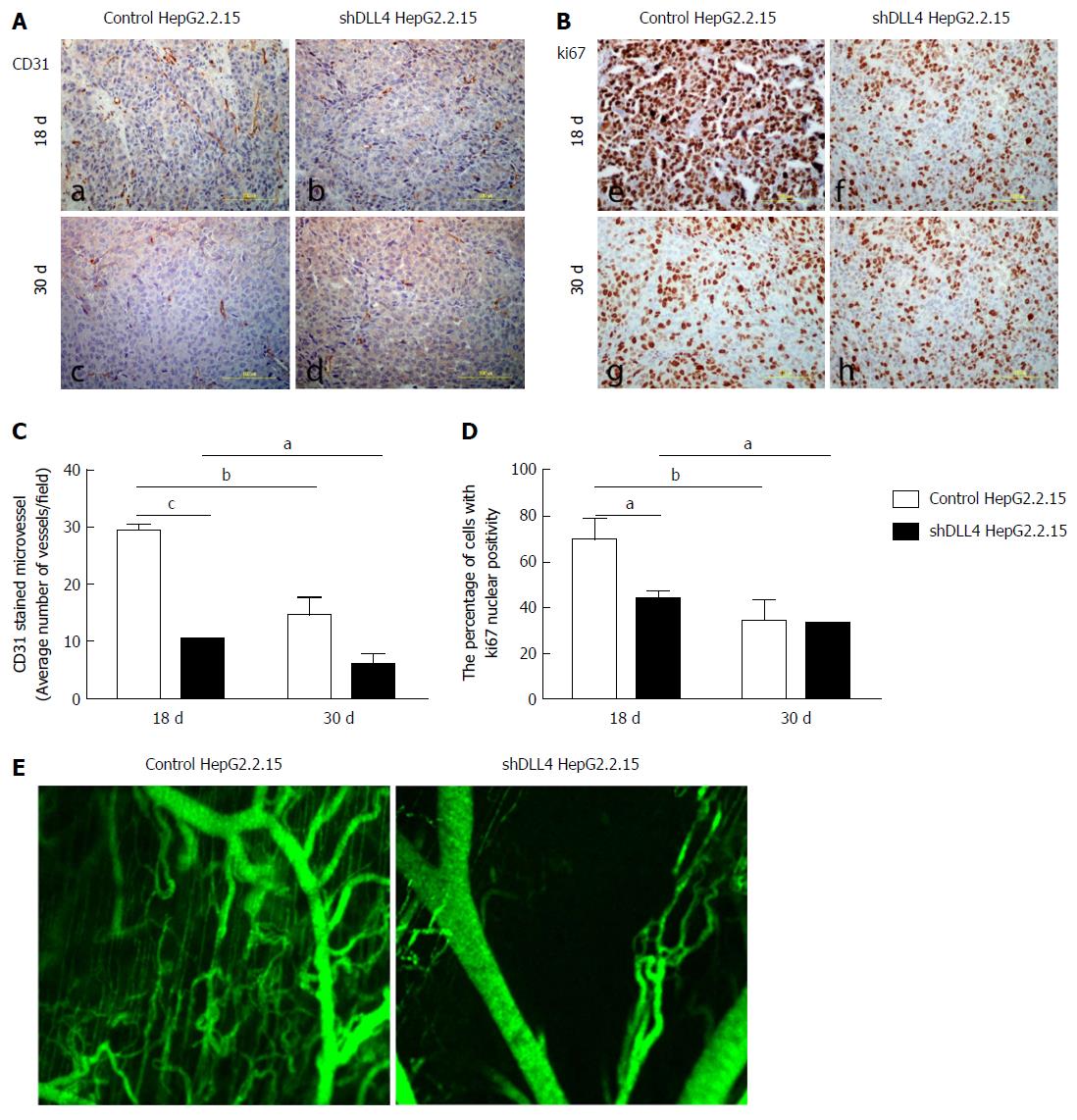
Figure 2 Suppression of Delta-like ligand 4 reduces tumour proliferation and neovasculature at the initiation stage of implantation.
A and B: Immunohistochemical staining of CD31 (a-d) and Ki67 (e-h) shows mouse neovessels and tumour cell proliferation in paraffin sections of tumour xenografts, respectively. Five fields of each section were quantified for the amount of CD31 staining (C), and the percentage of Ki67 positive cells (D) in tumours transfected with shDLL4 or control vector at 18 d and 30 d after implantation. The tumour vasculature was measured after tumour implantation with shDLL4 HepG2.2.15 or control cells at 30 d (E). The data represent the mean ± SD. aP < 0.05; bP < 0.01; cP < 0.001.
- Citation: Kunanopparat A, Issara-Amphorn J, Leelahavanichkul A, Sanpavat A, Patumraj S, Tangkijvanich P, Palaga T, Hirankarn N. Delta-like ligand 4 in hepatocellular carcinoma intrinsically promotes tumour growth and suppresses hepatitis B virus replication. World J Gastroenterol 2018; 24(34): 3861-3870
- URL: https://www.wjgnet.com/1007-9327/full/v24/i34/3861.htm
- DOI: https://dx.doi.org/10.3748/wjg.v24.i34.3861


