Copyright
©The Author(s) 2018.
World J Gastroenterol. Aug 21, 2018; 24(31): 3556-3566
Published online Aug 21, 2018. doi: 10.3748/wjg.v24.i31.3556
Published online Aug 21, 2018. doi: 10.3748/wjg.v24.i31.3556
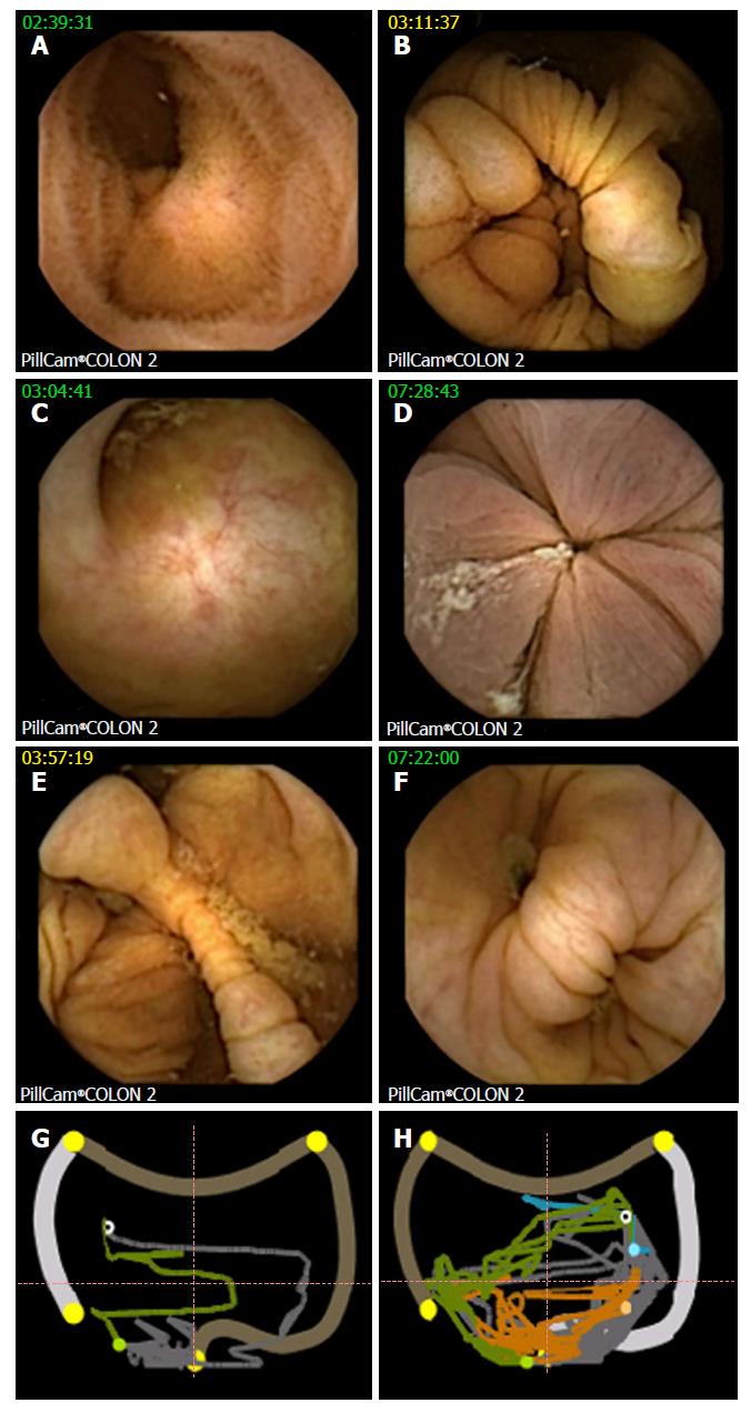
Figure 1 Landmarks at colon capsule endoscopy.
A: Terminal ileum; B: IC valve; C: Appendix; D: Hemorrhoidal plexus, hepatic flexure - CCE image (E) with corresponding localization trace (G), white circle showing actual capsule position, green colon already displayed, grey colon yet to be analyzed, orange small bowel, blue stomach, outer pictogram the position to the colonic segment as manually defined by setting the landmarks; F and H: CCE image and localization trace of splenic flexure in another patient. CCE: Colon capsule endoscopy.
- Citation: Baltes P, Bota M, Albert J, Philipper M, Hörster HG, Hagenmüller F, Steinbrück I, Jakobs R, Bechtler M, Hartmann D, Neuhaus H, Charton JP, Mayershofer R, Hohn H, Rösch T, Groth S, Nowak T, Wohlmuth P, Keuchel M. PillCamColon2 after incomplete colonoscopy - A prospective multicenter study. World J Gastroenterol 2018; 24(31): 3556-3566
- URL: https://www.wjgnet.com/1007-9327/full/v24/i31/3556.htm
- DOI: https://dx.doi.org/10.3748/wjg.v24.i31.3556


