Copyright
©The Author(s) 2018.
World J Gastroenterol. Jan 21, 2018; 24(3): 360-370
Published online Jan 21, 2018. doi: 10.3748/wjg.v24.i3.360
Published online Jan 21, 2018. doi: 10.3748/wjg.v24.i3.360
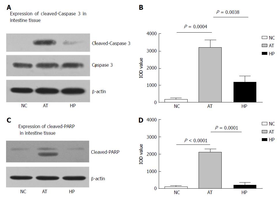
Figure 4 Changes of Caspase 3 and PARP expression in rat intestinal tissue at postoperative 24 h.
A: The apoptosis was detected by Western blot assay, which showed that the expression of cleaved Caspase 3 as increased in rat intestinal tissue with ischemia and reperfusion injury at postoperative 24 h; B: The bar-table shows the level of cleaved Caspase 3 expression in AT group was significantly higher than that in the NC group. The cleaved Caspase 3 expression in the HP group was lower than that in the AT group at postoperative 24 h; C: The expression of cleaved PARP was increased in rat intestinal tissue with ischemia and reperfusion injury at postoperative 24 h; D: The bar-table shows that the level of cleaved PARP expression in the AT group was significantly higher than that in the NC group. The cleaved PARP expression in the HP group was lower than that in the AT group at postoperative 24 h.
- Citation: Ji ZP, Li YX, Shi BX, Zhuang ZN, Yang JY, Guo S, Xu XZ, Xu KS, Li HL. Hypoxia preconditioning protects Ca2+-ATPase activation of intestinal mucosal cells against R/I injury in a rat liver transplantation model. World J Gastroenterol 2018; 24(3): 360-370
- URL: https://www.wjgnet.com/1007-9327/full/v24/i3/360.htm
- DOI: https://dx.doi.org/10.3748/wjg.v24.i3.360


