Copyright
©The Author(s) 2018.
World J Gastroenterol. Aug 7, 2018; 24(29): 3273-3280
Published online Aug 7, 2018. doi: 10.3748/wjg.v24.i29.3273
Published online Aug 7, 2018. doi: 10.3748/wjg.v24.i29.3273
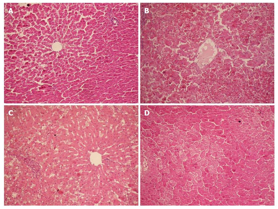
Figure 1 Histopathology of the liver (HE staining, × 200).
A: Group Normal Saline (NS). Normal hepatocytes were arranged in cords; B: Group D-galactosamine (D-GalN) plus lipopolysaccharide (LPS) (G/L). At 12 h, massive hepatocyte necrosis with severe hemorrhage developed; C: Group D-GaIN (G). At 12 h, spotty hepatocyte necrosis was observed; C: Group LPS (L). At 12 h, hepatocytes began to develop necrosis, with incomplete necrosis visible.
- Citation: Wang JB, Gu Y, Zhang MX, Yang S, Wang Y, Wang W, Li XR, Zhao YT, Wang HT. High expression of type I inositol 1,4,5-trisphosphate receptor in the kidney of rats with hepatorenal syndrome. World J Gastroenterol 2018; 24(29): 3273-3280
- URL: https://www.wjgnet.com/1007-9327/full/v24/i29/3273.htm
- DOI: https://dx.doi.org/10.3748/wjg.v24.i29.3273


