Copyright
©The Author(s) 2018.
World J Gastroenterol. Aug 7, 2018; 24(29): 3250-3259
Published online Aug 7, 2018. doi: 10.3748/wjg.v24.i29.3250
Published online Aug 7, 2018. doi: 10.3748/wjg.v24.i29.3250
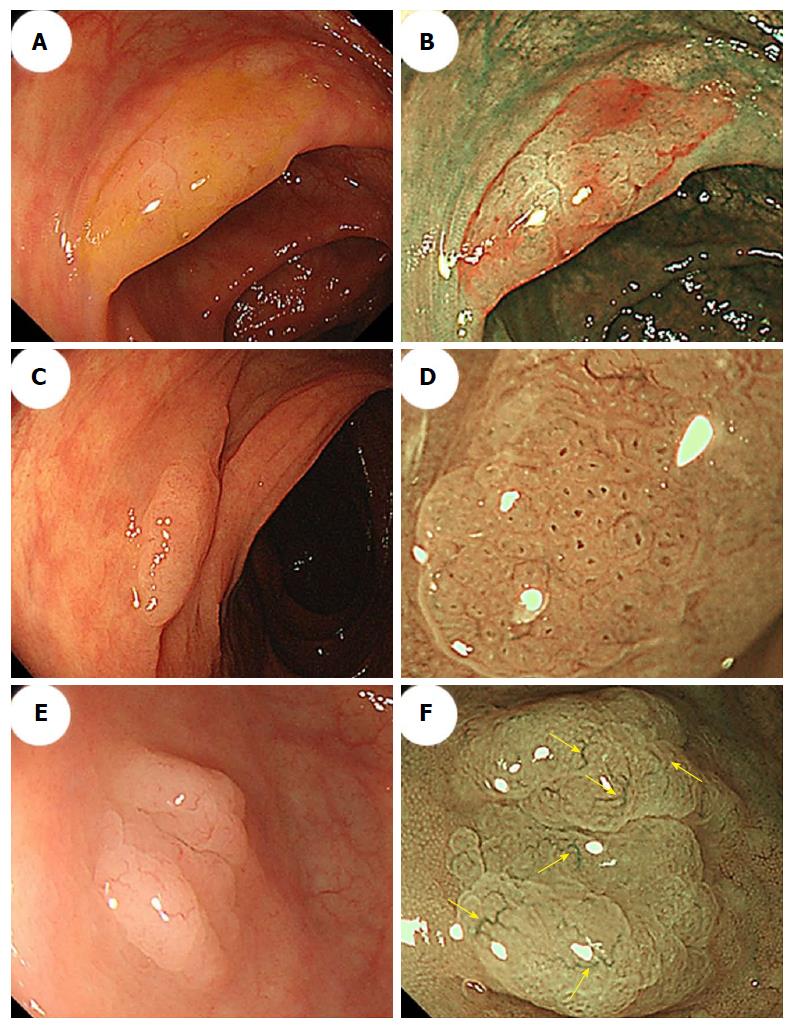
Figure 3 Morphologic characteristics of sessile serrated adenoma/polyps.
A: Conventional endoscopy revealed a flat-elevated lesion with a 20-mm diameter that was covered with a mucus cap in the transverse colon. B: Narrow-band imaging (NBI) showed that the SSA/P in (A) was covered with a mucus cap that appeared intensely red. C: Conventional endoscopy showed a flat-elevated lesion with a 14-mm diameter in the ascending colon. D: Magnifying NBI of the SSA/P in (C) revealed dark spots inside the crypts in part of the lesion. E: A conventional endoscopic image shows a flat-elevated pale colored lesion with a 10-mm diameter in the cecum. F: Magnifying NBI of the SSA/P in (E) revealed varicose microvascular vessels (arrows) in part of the lesion. SSA/P: Sessile serrated adenoma/polyp.
- Citation: Murakami T, Sakamoto N, Nagahara A. Endoscopic diagnosis of sessile serrated adenoma/polyp with and without dysplasia/carcinoma. World J Gastroenterol 2018; 24(29): 3250-3259
- URL: https://www.wjgnet.com/1007-9327/full/v24/i29/3250.htm
- DOI: https://dx.doi.org/10.3748/wjg.v24.i29.3250


