Copyright
©The Author(s) 2018.
World J Gastroenterol. Jul 21, 2018; 24(27): 3030-3037
Published online Jul 21, 2018. doi: 10.3748/wjg.v24.i27.3030
Published online Jul 21, 2018. doi: 10.3748/wjg.v24.i27.3030
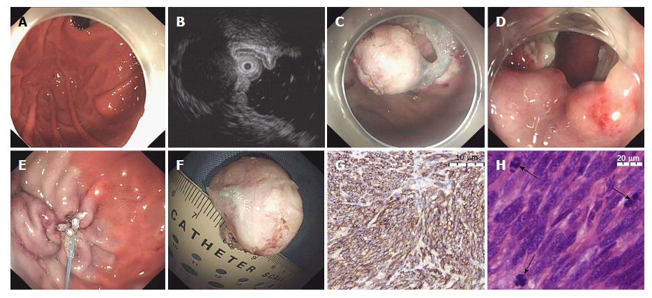
Figure 1 Endoscopic full-thickness resection for a gastrointestinal stromal tumors located in the gastric fundus.
A: Endoscopy showed a SET was located in the gastric fundus; B: EUS showed the same tumor mainly bulged into the extraluminal space and had an extensive connection to the MP layer of the stomach; C: The tumor, including its underlying MP and serosa, was resected; D and E: The gastric wall defect was closed using clips combined with an endoloop; F: The resection specimen was a 2.7 cm tumor; G: Immunohistochemistry showed that CD117 was present in most tumor cells (original magnification × 100); H: Mitotic figures could be found easily (original magnification × 400). EUS: Endoscopic ultrasonography.
- Citation: Zhang Y, Mao XL, Zhou XB, Yang H, Zhu LH, Chen G, Ye LP. Long-term outcomes of endoscopic resection for small (≤ 4.0 cm) gastric gastrointestinal stromal tumors originating from the muscularis propria layer. World J Gastroenterol 2018; 24(27): 3030-3037
- URL: https://www.wjgnet.com/1007-9327/full/v24/i27/3030.htm
- DOI: https://dx.doi.org/10.3748/wjg.v24.i27.3030


