Copyright
©The Author(s) 2018.
World J Gastroenterol. Jul 21, 2018; 24(27): 2921-2930
Published online Jul 21, 2018. doi: 10.3748/wjg.v24.i27.2921
Published online Jul 21, 2018. doi: 10.3748/wjg.v24.i27.2921
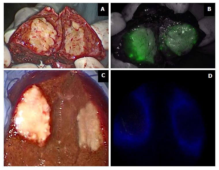
Figure 3 Indocyanine green in liver surgery.
Primary liver tumors show intense and complete staining because their hepatocytes take up ICG but do not secrete it (A and B); liver metastases show a ring appearance because their cells do not take up ICG but hepatocytes surrounding the nodule are compressed (C and D). ICG: Indocyanine green.
- Citation: Baiocchi GL, Diana M, Boni L. Indocyanine green-based fluorescence imaging in visceral and hepatobiliary and pancreatic surgery: State of the art and future directions. World J Gastroenterol 2018; 24(27): 2921-2930
- URL: https://www.wjgnet.com/1007-9327/full/v24/i27/2921.htm
- DOI: https://dx.doi.org/10.3748/wjg.v24.i27.2921


