Copyright
©The Author(s) 2018.
World J Gastroenterol. Jul 7, 2018; 24(25): 2776-2784
Published online Jul 7, 2018. doi: 10.3748/wjg.v24.i25.2776
Published online Jul 7, 2018. doi: 10.3748/wjg.v24.i25.2776
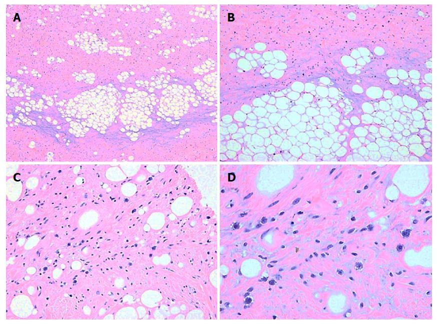
Figure 1 Histological finding.
Low magnification shows that the immature fat cells are interspersed among smooth muscle tissues (A: HE, × 40 and B: HE; × 100). The large nuclear dark-stained lipoblast, which appears as a mononuclear or multinucleated cell with one or more cytoplasmic vacuoles, are seen under high magnification (C; HE; × 200 and D: HE; × 400).
- Citation: Kang WZ, Xue LY, Wang GQ, Ma FH, Feng XL, Guo L, Li Y, Li WK, Tian YT. Liposarcoma of the stomach: Report of two cases and review of the literature. World J Gastroenterol 2018; 24(25): 2776-2784
- URL: https://www.wjgnet.com/1007-9327/full/v24/i25/2776.htm
- DOI: https://dx.doi.org/10.3748/wjg.v24.i25.2776


