Copyright
©The Author(s) 2018.
World J Gastroenterol. Jun 28, 2018; 24(24): 2640-2646
Published online Jun 28, 2018. doi: 10.3748/wjg.v24.i24.2640
Published online Jun 28, 2018. doi: 10.3748/wjg.v24.i24.2640
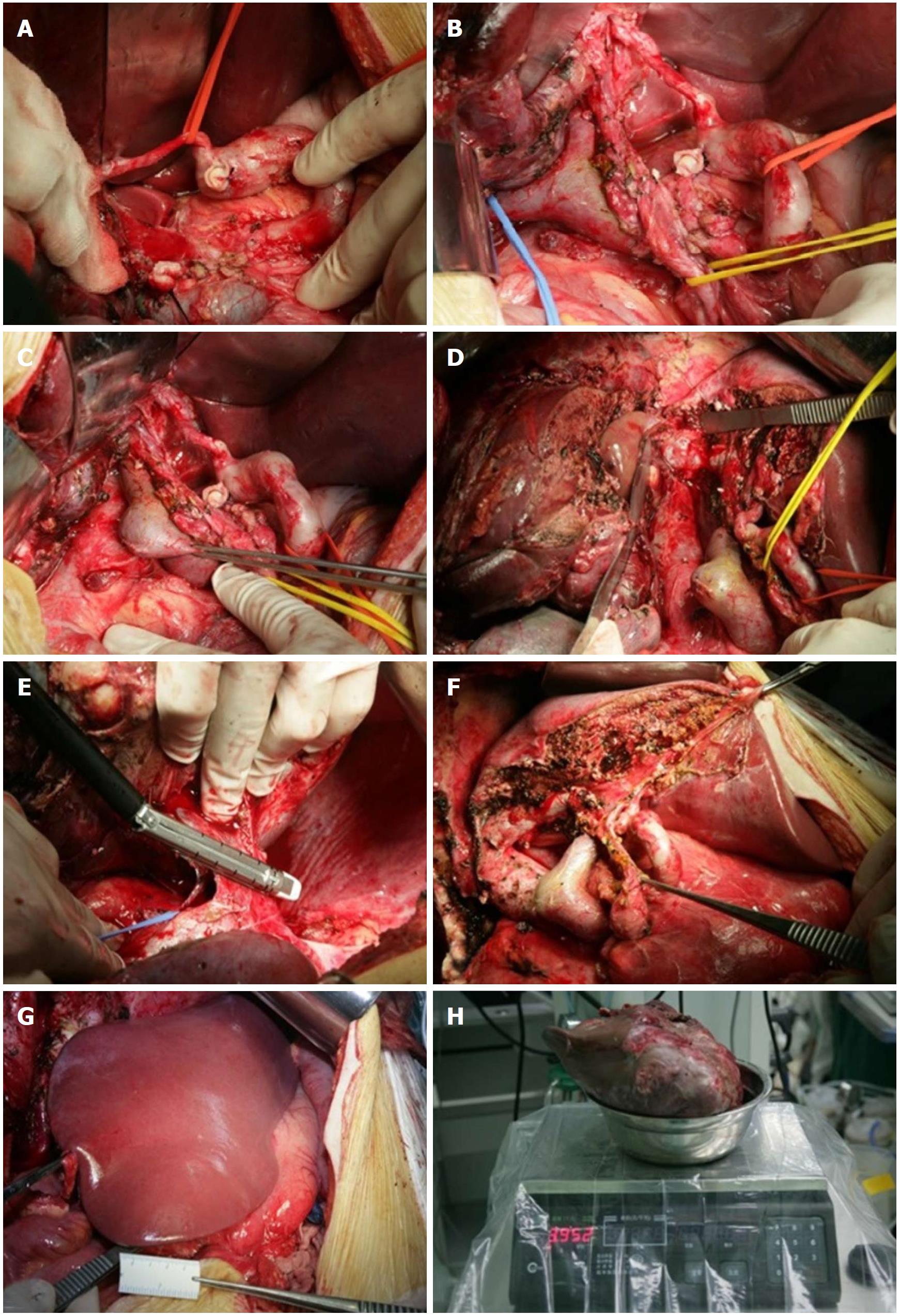
Figure 4 Surgical procedures.
A: Separation of the hepatic artery, suspension of the left hepatic artery, and the right hepatic artery was ligated and transected; B: Separation of the portal vein and bile duct. The right hepatic duct was transected and sutured; C: Separation of the right portal vein; D: Splitting the liver parenchyma to the inferior vena cava; E: Severing of the right hepatic vein using the stapler; F: Condition after surgical resection, showing that the left hepatic artery, left portal vein, and left hepatic duct were structurally intact; G: The remaining liver after resection was ruddy in color, with no ischemia or blood stasis; H: The specimen weight was 3952 g.
- Citation: Meng XF, Pan YW, Wang ZB, Duan WD. Primary hepatic neuroendocrine tumor case with a preoperative course of 26 years: A case report and literature review. World J Gastroenterol 2018; 24(24): 2640-2646
- URL: https://www.wjgnet.com/1007-9327/full/v24/i24/2640.htm
- DOI: https://dx.doi.org/10.3748/wjg.v24.i24.2640


