Copyright
©The Author(s) 2018.
World J Gastroenterol. Jun 14, 2018; 24(22): 2406-2412
Published online Jun 14, 2018. doi: 10.3748/wjg.v24.i22.2406
Published online Jun 14, 2018. doi: 10.3748/wjg.v24.i22.2406
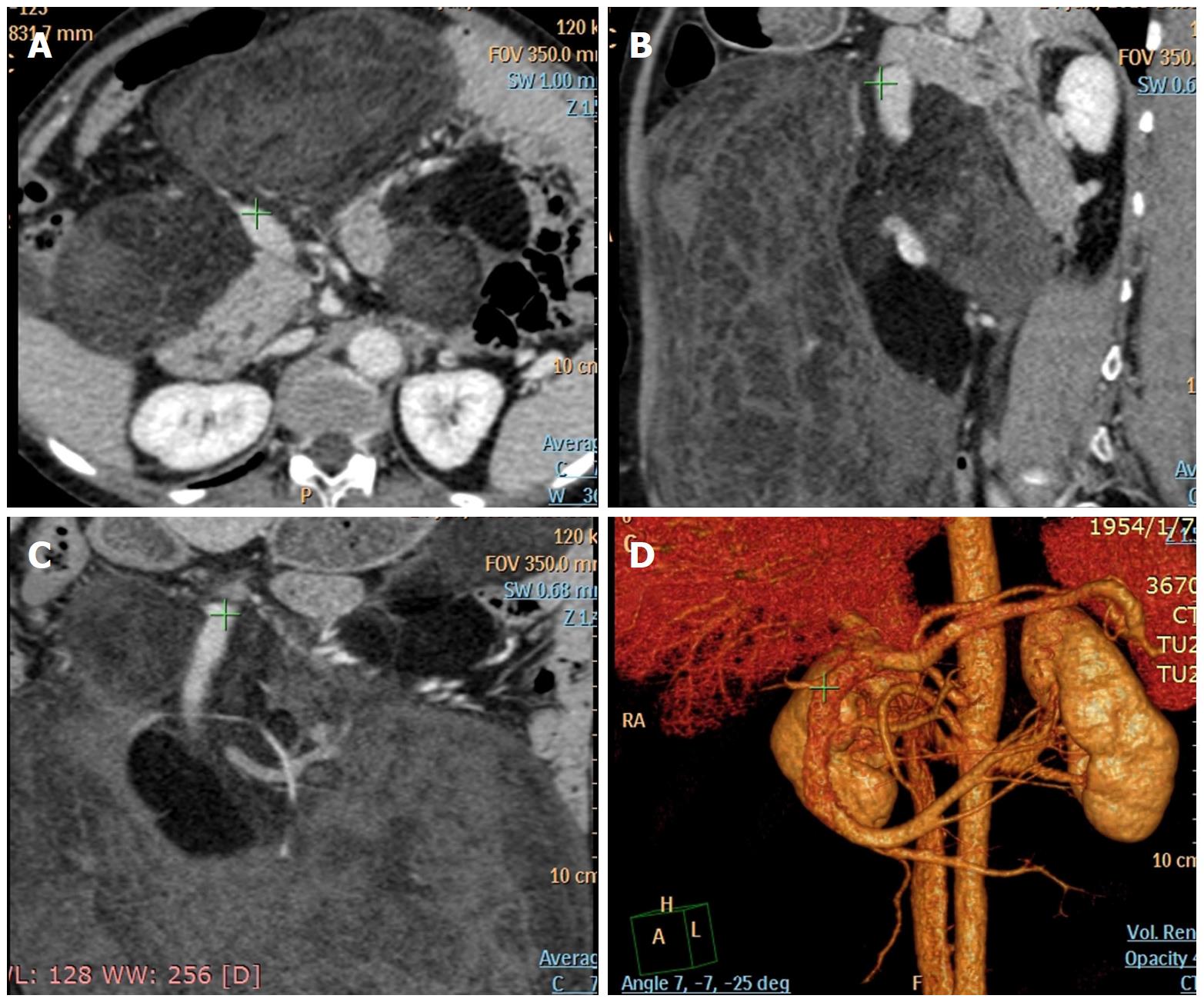
Figure 2 Preoperative abdominal computed tomographic angiography.
A-C: The same location (green cross) of the main trunk of the superior mesenteric vein in the horizontal scan, coronary scan, and sagittal scan. D: 3-dimensional CT scan showing that the lesion adhered to and constricted the main trunk of the superior mesenteric vein.
- Citation: Miao RC, Wan Y, Zhang XG, Zhang X, Deng Y, Liu C. Devascularization of the superior mesenteric vein without reconstruction during surgery for retroperitoneal liposarcoma: A case report and review of literature. World J Gastroenterol 2018; 24(22): 2406-2412
- URL: https://www.wjgnet.com/1007-9327/full/v24/i22/2406.htm
- DOI: https://dx.doi.org/10.3748/wjg.v24.i22.2406


