Copyright
©The Author(s) 2018.
World J Gastroenterol. Jun 14, 2018; 24(22): 2348-2362
Published online Jun 14, 2018. doi: 10.3748/wjg.v24.i22.2348
Published online Jun 14, 2018. doi: 10.3748/wjg.v24.i22.2348
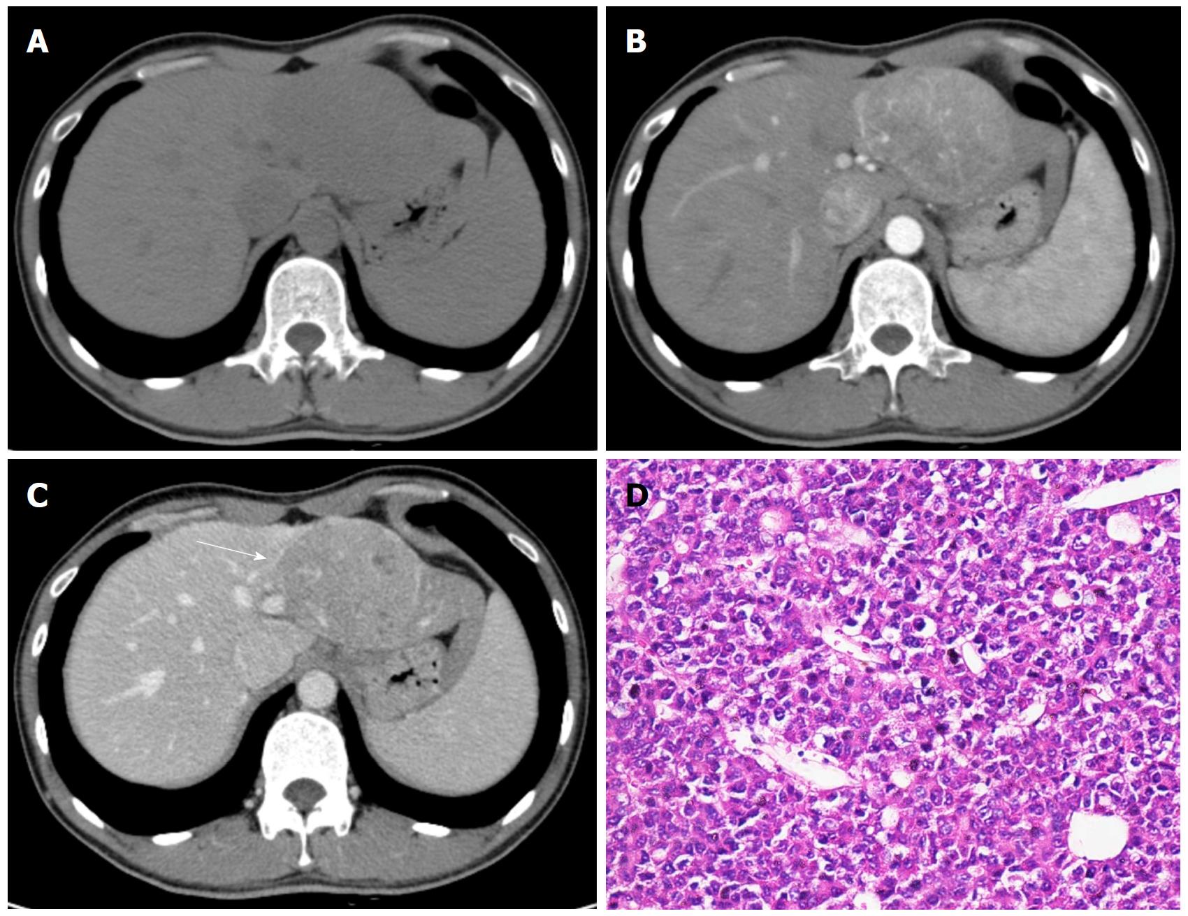
Figure 1 Hepatocellular carcinoma in a 32-year-old male with chronic hepatitis B.
Axial dynamic non-enhanced (A), late arterial phase (B), and portal venous phase (C) CT images show the 8.5 cm mass with arterial phase hyperenhancement and portal venous phase wash-out appearance. The capsule is seen as a hyperattenuating ring on portal venous phase (C, white arrow). The hematoxylin-eosin (HE) staining of the mass at 200 × magnification proved it to be Edmonson-Steiner grade II (D).
- Citation: Jiang HY, Chen J, Xia CC, Cao LK, Duan T, Song B. Noninvasive imaging of hepatocellular carcinoma: From diagnosis to prognosis. World J Gastroenterol 2018; 24(22): 2348-2362
- URL: https://www.wjgnet.com/1007-9327/full/v24/i22/2348.htm
- DOI: https://dx.doi.org/10.3748/wjg.v24.i22.2348


