Copyright
©The Author(s) 2018.
World J Gastroenterol. Jun 7, 2018; 24(21): 2279-2290
Published online Jun 7, 2018. doi: 10.3748/wjg.v24.i21.2279
Published online Jun 7, 2018. doi: 10.3748/wjg.v24.i21.2279
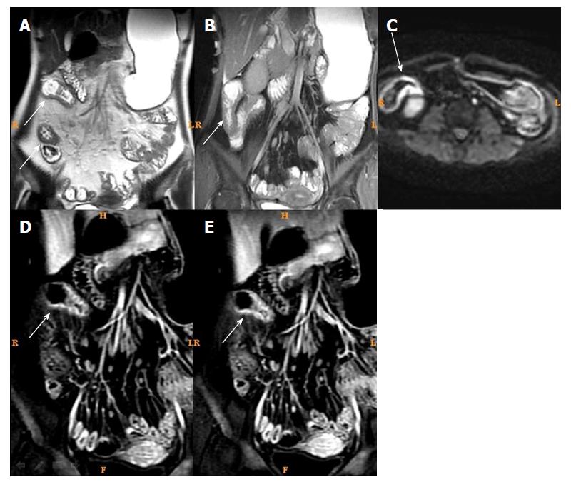
Figure 1 Magnetic resonance imaging of a typical case of active Crohn’s disease before treatment.
Female, 32 years of age, active Crohn’s disease. A: T2WI showed intestinal wall thickening and submucosal edema in the distal ileum; B: Fast imaging employing steady-state acquisition showed intestinal wall thickening and submucosal edema in the distal ileum; C: Diffusion weight imaging showed marked high intensity; D and E: Dynamic enhancement showed obvious layer stratified enhancement.
- Citation: Zhu NY, Zhao XS, Miao F. Magnetic resonance imaging and Crohn’s disease endoscopic index of severity: Correlations and concordance. World J Gastroenterol 2018; 24(21): 2279-2290
- URL: https://www.wjgnet.com/1007-9327/full/v24/i21/2279.htm
- DOI: https://dx.doi.org/10.3748/wjg.v24.i21.2279


