Copyright
©The Author(s) 2018.
World J Gastroenterol. Jan 14, 2018; 24(2): 297-302
Published online Jan 14, 2018. doi: 10.3748/wjg.v24.i2.297
Published online Jan 14, 2018. doi: 10.3748/wjg.v24.i2.297
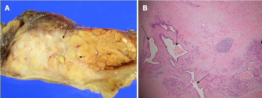
Figure 4 Macroscopic and microscopic findings of resected specimen.
A: Gross specimen shows whitish hard infiltrating mass-like lesion (arrows) focally replaced head and uncinate process of pancreas; B: Microscopy (hematoxylin and eosin, x 40) shows perilobular and intralobular fibrosis (asterisk) replaces normal pancreatic acini with focal perivascular lymphocyte infiltration (short arrow) and dilated branch ducts (long arrows).
- Citation: Jee KN. Mass forming chronic pancreatitis mimicking pancreatic cystic neoplasm: A case report. World J Gastroenterol 2018; 24(2): 297-302
- URL: https://www.wjgnet.com/1007-9327/full/v24/i2/297.htm
- DOI: https://dx.doi.org/10.3748/wjg.v24.i2.297


