Copyright
©The Author(s) 2018.
World J Gastroenterol. Jan 14, 2018; 24(2): 195-210
Published online Jan 14, 2018. doi: 10.3748/wjg.v24.i2.195
Published online Jan 14, 2018. doi: 10.3748/wjg.v24.i2.195
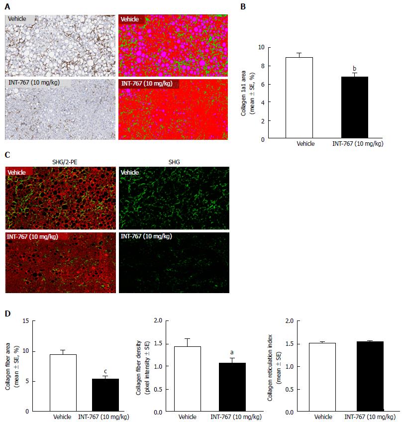
Figure 2 INT-767 treatment for 8 wk reduces hepatic collagen deposition in ob/ob-NASH mice with biopsy-confirmed liver pathology.
A: Collagen 1a1 (immunohistochemistry); B: Fractional area of collagen (immunohistochemistry); C: Label free SHG/2-PE images for collagen fiber deposition (green) in hepatic parenchyma (red). Data are expressed as % of total parenchymal area (subtraction of fat area); D: Fractional area of collagen fiber, collagen fiber density, and collagen fiber reticulation index (SHG analysis). aP < 0.05, bP < 0.01, cP < 0.001 (unpaired t-test).
- Citation: Roth JD, Feigh M, Veidal SS, Fensholdt LK, Rigbolt KT, Hansen HH, Chen LC, Petitjean M, Friley W, Vrang N, Jelsing J, Young M. INT-767 improves histopathological features in a diet-induced ob/ob mouse model of biopsy-confirmed non-alcoholic steatohepatitis. World J Gastroenterol 2018; 24(2): 195-210
- URL: https://www.wjgnet.com/1007-9327/full/v24/i2/195.htm
- DOI: https://dx.doi.org/10.3748/wjg.v24.i2.195


