Copyright
©The Author(s) 2018.
World J Gastroenterol. May 21, 2018; 24(19): 2130-2136
Published online May 21, 2018. doi: 10.3748/wjg.v24.i19.2130
Published online May 21, 2018. doi: 10.3748/wjg.v24.i19.2130
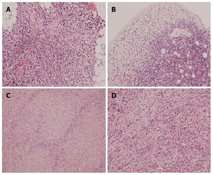
Figure 5 Histopathologic evaluation performed after open D2 gastrectomy combined with liver metastasectomy.
A: Tumor microfocus infiltrations in the peritoneal adipose tissue in the vicinity of the distal surgical margin (obj. 20 ×, HE); B: Metastatic foci in the subcapsular region of the lymph node (obj. 20 ×, HE); C, D: Liver metastasis of gastric carcinoma (obj. 10 ×, 20 ×, HE).
- Citation: Nowacki M, Grzanka D, Zegarski W. Pressurized intraperitoneal aerosol chemotheprapy after misdiagnosed gastric cancer: Case report and review of the literature. World J Gastroenterol 2018; 24(19): 2130-2136
- URL: https://www.wjgnet.com/1007-9327/full/v24/i19/2130.htm
- DOI: https://dx.doi.org/10.3748/wjg.v24.i19.2130


