Copyright
©The Author(s) 2018.
World J Gastroenterol. May 21, 2018; 24(19): 2130-2136
Published online May 21, 2018. doi: 10.3748/wjg.v24.i19.2130
Published online May 21, 2018. doi: 10.3748/wjg.v24.i19.2130
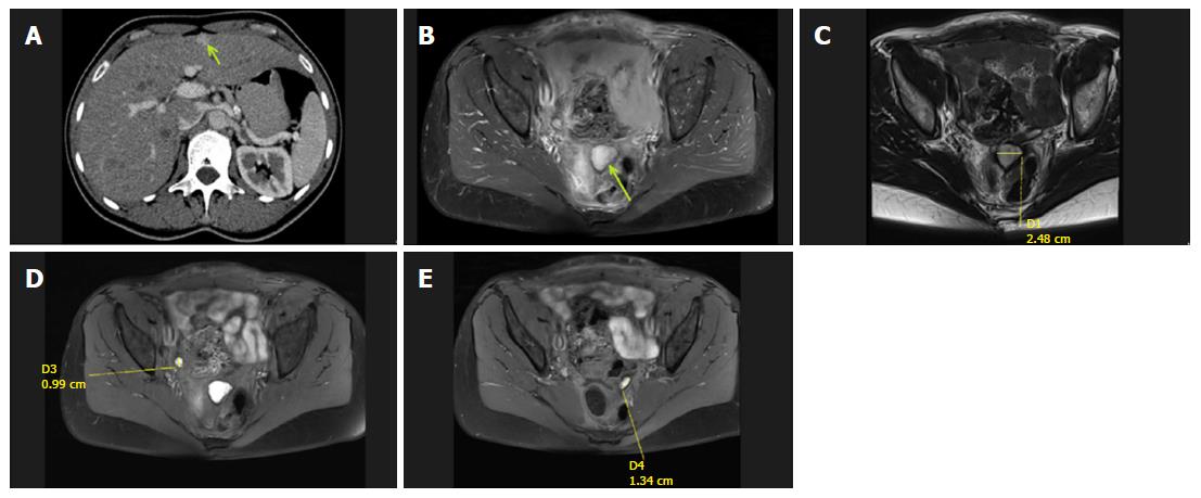
Figure 2 Abdominal computed tomography and abdominopelvic magnetic resonance scans.
A: Post-contrast computed tomography image in the portal venous phase showing a hyperintense enhancing lesion in segment V (isodense in the native phase) of the liver, which was diagnosed as a superficially located suspicious metastatic lesion; B-E: Abdominopelvic magnetic resonance scans with evident masses and suspicious nodules.
- Citation: Nowacki M, Grzanka D, Zegarski W. Pressurized intraperitoneal aerosol chemotheprapy after misdiagnosed gastric cancer: Case report and review of the literature. World J Gastroenterol 2018; 24(19): 2130-2136
- URL: https://www.wjgnet.com/1007-9327/full/v24/i19/2130.htm
- DOI: https://dx.doi.org/10.3748/wjg.v24.i19.2130


