Copyright
©The Author(s) 2018.
World J Gastroenterol. May 21, 2018; 24(19): 2130-2136
Published online May 21, 2018. doi: 10.3748/wjg.v24.i19.2130
Published online May 21, 2018. doi: 10.3748/wjg.v24.i19.2130
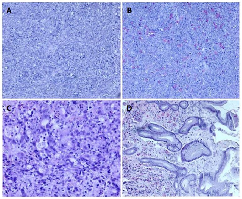
Figure 1 Histological ovary assay.
A: Signet ring of tumor cell infiltration within the ovarian stroma (10 ×, HE); B: Mucicarmine staining highlights the presence of mucin in the cytoplasm (10 ×). Histopathologic evaluation of antral mucosa showing (C) poorly differentiated carcinoma infiltration with signet ring cells (20 ×, HE) and (D) mucicarmine staining of mucin-positive cells in the gastric mucosa (10 ×).
- Citation: Nowacki M, Grzanka D, Zegarski W. Pressurized intraperitoneal aerosol chemotheprapy after misdiagnosed gastric cancer: Case report and review of the literature. World J Gastroenterol 2018; 24(19): 2130-2136
- URL: https://www.wjgnet.com/1007-9327/full/v24/i19/2130.htm
- DOI: https://dx.doi.org/10.3748/wjg.v24.i19.2130


