Copyright
©The Author(s) 2018.
World J Gastroenterol. May 14, 2018; 24(18): 1925-1941
Published online May 14, 2018. doi: 10.3748/wjg.v24.i18.1925
Published online May 14, 2018. doi: 10.3748/wjg.v24.i18.1925
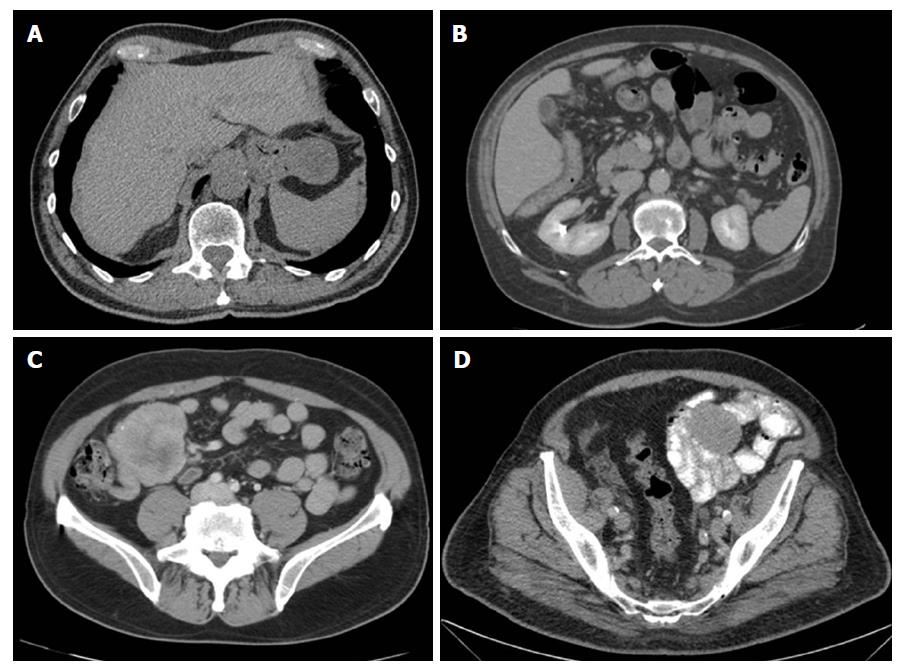
Figure 1 Localized gastrointestinal stromal tumors on computed tomography scan.
A: Gastric; B: Duodenal; C: Ileal; D: Jejunal. A and B show respectively a gastric tumor and a duodenal tumor of exophytic growth with well-defined borders. Appreciate in C the different densities inside the tumor due to due to necrosis, haemorrhage, or degenerative components. D shows a jejunal GIST in left iliac fossa.
- Citation: Sanchez-Hidalgo JM, Duran-Martinez M, Molero-Payan R, Rufian-Peña S, Arjona-Sanchez A, Casado-Adam A, Cosano-Alvarez A, Briceño-Delgado J. Gastrointestinal stromal tumors: A multidisciplinary challenge. World J Gastroenterol 2018; 24(18): 1925-1941
- URL: https://www.wjgnet.com/1007-9327/full/v24/i18/1925.htm
- DOI: https://dx.doi.org/10.3748/wjg.v24.i18.1925


