Copyright
©The Author(s) 2018.
World J Gastroenterol. May 7, 2018; 24(17): 1888-1900
Published online May 7, 2018. doi: 10.3748/wjg.v24.i17.1888
Published online May 7, 2018. doi: 10.3748/wjg.v24.i17.1888
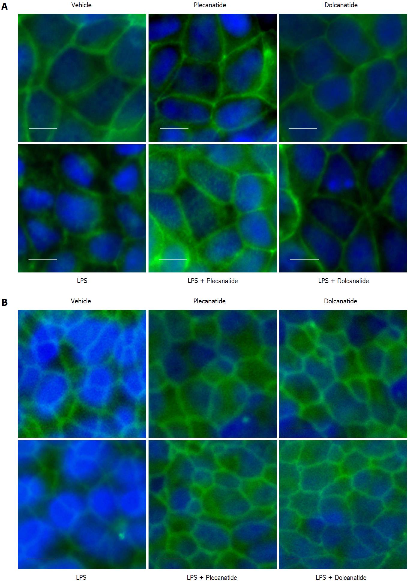
Figure 4 Effect of plecanatide and dolcanatide on localization of ZO-1 in epithelial cells Caco-2 (A) and T84 (B) cell monolayers were treated with 1 μmol/L plecanatide or dolcanatide in the presence of 100 μg/mL of LPS for 16 h followed by immunofluorescence imaging for ZO-1.
Representative microscopic fields depicted above demonstrate disruption of ZO-1 localization by LPS. In Caco-2 cells, LPS treatment appears to cause accumulation of ZO-1 in the cytoplasm. Co-treatment of LPS with plecanatide or dolcanatide preserved ZO-1 localization around the cell membrane as observed for vehicle treated cells. Images taken at 40× resolution. Blue fluorescence corresponds to DAPI stained nucleus. DAPI: 4’, 6’-Diamidino-2-phenylindole; LPS: Lipopolysaccharide; ZO-1: Zonula occludens-1.
- Citation: Boulete IM, Thadi A, Beaufrand C, Patwa V, Joshi A, Foss JA, Eddy EP, Eutamene H, Palejwala VA, Theodorou V, Shailubhai K. Oral treatment with plecanatide or dolcanatide attenuates visceral hypersensitivity via activation of guanylate cyclase-C in rat models. World J Gastroenterol 2018; 24(17): 1888-1900
- URL: https://www.wjgnet.com/1007-9327/full/v24/i17/1888.htm
- DOI: https://dx.doi.org/10.3748/wjg.v24.i17.1888


