Copyright
©The Author(s) 2018.
World J Gastroenterol. Apr 28, 2018; 24(16): 1779-1794
Published online Apr 28, 2018. doi: 10.3748/wjg.v24.i16.1779
Published online Apr 28, 2018. doi: 10.3748/wjg.v24.i16.1779
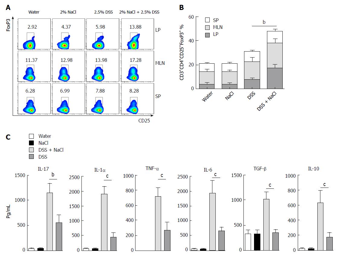
Figure 4 CD3+CD4+CD25+Foxp3+ T cells are increased in mice treated with DSS and NaCl.
A: CD3+CD4+CD25+Foxp3+ T cells in LP, MLN and SP from animal models were detected by flow cytometry; B: A summary of the percentages of CD3+CD4+CD25+Foxp3+ T cell distribution in LP, MLN and SP; C: LPMCs from the four groups were isolated and cultured for 24 h, and the levels of cytokines in the culture supernatants were collected and analyzed by enzyme-linked immunosorbent assay. In all the panels, data indicate three separate experiments, whereby 3 mice per group were used in each experiment. aP < 0.05; bP < 0.01; cP < 0.001 vs the DSS group. DSS: Dextran sulfate sodium; LMPCs: Lamina propria mononuclear cells; LP: Lamina propria; MLN: Mesenteric lymph node; SP: Spleen.
- Citation: Guo HX, Ye N, Yan P, Qiu MY, Zhang J, Shen ZG, He HY, Tian ZQ, Li HL, Li JT. Sodium chloride exacerbates dextran sulfate sodium-induced colitis by tuning proinflammatory and antiinflammatory lamina propria mononuclear cells through p38/MAPK pathway in mice. World J Gastroenterol 2018; 24(16): 1779-1794
- URL: https://www.wjgnet.com/1007-9327/full/v24/i16/1779.htm
- DOI: https://dx.doi.org/10.3748/wjg.v24.i16.1779


