Copyright
©The Author(s) 2017.
World J Gastroenterol. Mar 7, 2017; 23(9): 1725-1734
Published online Mar 7, 2017. doi: 10.3748/wjg.v23.i9.1725
Published online Mar 7, 2017. doi: 10.3748/wjg.v23.i9.1725
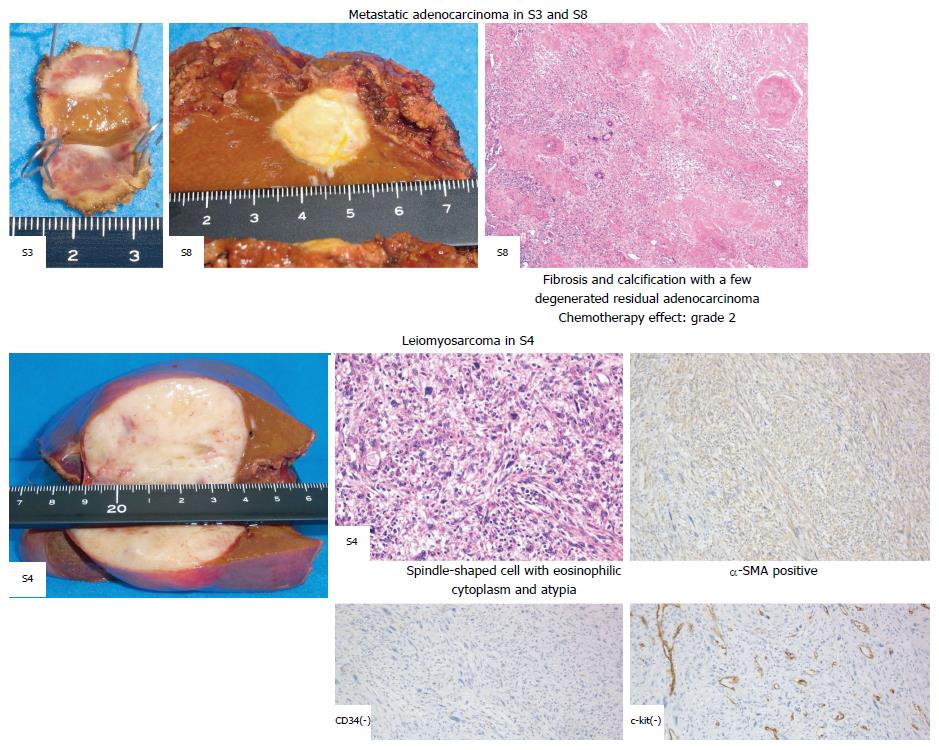
Figure 4 Pathological diagnoses of live lesions.
The tumors in Segments 3 and 8 revealed fibrosis and calcification, with a few degenerated residual adenocarcinomas, while the tumor in Segment 4 consisted of irregular fascicles of spindle-shaped cells and was positive for SMA and negative for CD34 and c-kit. SMA: Smooth muscle actin.
- Citation: Aoki H, Arata T, Utsumi M, Mushiake Y, Kunitomo T, Yasuhara I, Taniguchi F, Katsuda K, Tanakaya K, Takeuchi H, Yamasaki R. Synchronous coexistence of liver metastases from cecal leiomyosarcoma and rectal adenocarcinoma: A case report. World J Gastroenterol 2017; 23(9): 1725-1734
- URL: https://www.wjgnet.com/1007-9327/full/v23/i9/1725.htm
- DOI: https://dx.doi.org/10.3748/wjg.v23.i9.1725


