Copyright
©The Author(s) 2017.
World J Gastroenterol. Mar 7, 2017; 23(9): 1637-1644
Published online Mar 7, 2017. doi: 10.3748/wjg.v23.i9.1637
Published online Mar 7, 2017. doi: 10.3748/wjg.v23.i9.1637
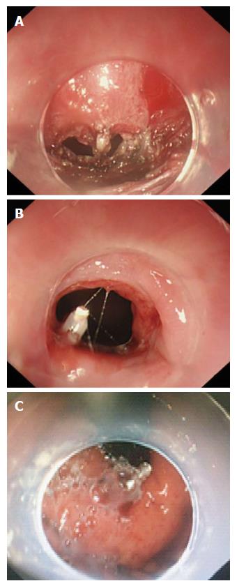
Figure 2 Closure of a 0.
8 cm × 0.4 cm mucosal penetration using a hemostatic clip and fibrin sealant. A: The appearance of the 0.8 cm × 0.4 cm mucosal penetration (imaging from the submucosal tunnel); B: A hemostatic clip was used to make a preliminary clipping (imaging from esophageal lumen); C: Fibrin sealant fully covers the preliminary clipped penetration (imaging from stomach lumen).
- Citation: Zhang WG, Linghu EQ, Li HK. Fibrin sealant for closure of mucosal penetration at the cardia during peroral endoscopic myotomy: A retrospective study at a single center. World J Gastroenterol 2017; 23(9): 1637-1644
- URL: https://www.wjgnet.com/1007-9327/full/v23/i9/1637.htm
- DOI: https://dx.doi.org/10.3748/wjg.v23.i9.1637


