Copyright
©The Author(s) 2017.
World J Gastroenterol. Feb 14, 2017; 23(6): 986-998
Published online Feb 14, 2017. doi: 10.3748/wjg.v23.i6.986
Published online Feb 14, 2017. doi: 10.3748/wjg.v23.i6.986
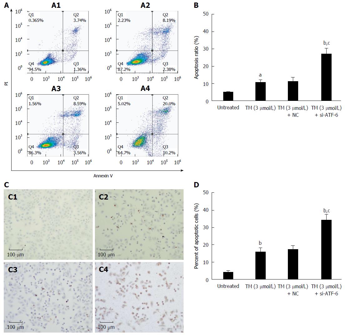
Figure 5 Effects of activating transcription factor 6 siRNA on cell apoptosis in HepG2 cells under endoplasmic reticulum stress.
A: HepG2 cells were transfected with ATF-6 siRNA for 24 h after pretreatment by tunicamycin (TM) for 8 h. Apoptotic cells were determined by FACS, and the data are expressed as the mean ± SD. A1, Untreated HepG2 cells; A2, HepG2 cells treated by TM only; A3, HepG2 cells treated by combination of TM and ATF-6 siRNA negative control; A4, HepG2 cells treated by ATF-6 siRNA and melatonin; B: Data are presented as the mean ± SD for the independent experiments (aP < 0.05 vs untreated HepG2 cells; bP < 0.01 vs untreated HepG2 cells; cP < 0.01 vs HepG2 cells treated with TM and ATF-6 siRNA negative control); C: Cell morphology and percentage of apoptotic cells was examined by TUNEL staining. C1, Untreated HepG2 cells; C2, HepG2 cells treated by TM only; C3, HepG2 cells treated by combination of TM and ATF-6 siRNA negative control; C4, HepG2 cells treated by ATF-6 siRNA and melatonin; D: Data are presented as the mean ± SD for the independent experiments (bP < 0.01 vs untreated HepG2 cells; cP < 0.01 vs HepG2 cells treated with TM and ATF-6 siRNA negative control). ATF-6: Activating transcription factor 6; NC: Negative control.
- Citation: Bu LJ, Yu HQ, Fan LL, Li XQ, Wang F, Liu JT, Zhong F, Zhang CJ, Wei W, Wang H, Sun GP. Melatonin, a novel selective ATF-6 inhibitor, induces human hepatoma cell apoptosis through COX-2 downregulation. World J Gastroenterol 2017; 23(6): 986-998
- URL: https://www.wjgnet.com/1007-9327/full/v23/i6/986.htm
- DOI: https://dx.doi.org/10.3748/wjg.v23.i6.986


