Copyright
©The Author(s) 2017.
World J Gastroenterol. Feb 14, 2017; 23(6): 1113-1118
Published online Feb 14, 2017. doi: 10.3748/wjg.v23.i6.1113
Published online Feb 14, 2017. doi: 10.3748/wjg.v23.i6.1113
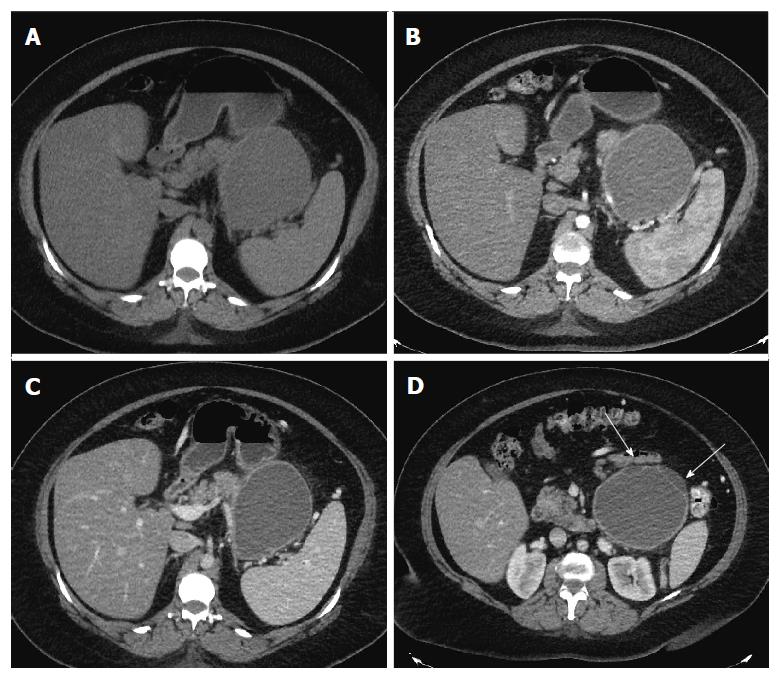
Figure 1 Axial computed tomography images.
Axial computed tomography (CT) images in unenhanced (A), pancreatic (B) and portal venous phase (C) show a well circumscribed, thin walled, large fluid density cystic lesion arising from and replacing the pancreatic body and tail, which abuts the spleen. The lesion does not exhibit post-contrast enhancement. Axial CT image at a lower level in portal venous phase (D) shows several thin septations and small loculations (arrows).
- Citation: Mederos MA, Villafañe N, Dhingra S, Farinas C, McElhany A, Fisher WE, Van Buren II G. Pancreatic endometrial cyst mimics mucinous cystic neoplasm of the pancreas. World J Gastroenterol 2017; 23(6): 1113-1118
- URL: https://www.wjgnet.com/1007-9327/full/v23/i6/1113.htm
- DOI: https://dx.doi.org/10.3748/wjg.v23.i6.1113


