Copyright
©The Author(s) 2017.
World J Gastroenterol. Feb 14, 2017; 23(6): 1106-1112
Published online Feb 14, 2017. doi: 10.3748/wjg.v23.i6.1106
Published online Feb 14, 2017. doi: 10.3748/wjg.v23.i6.1106
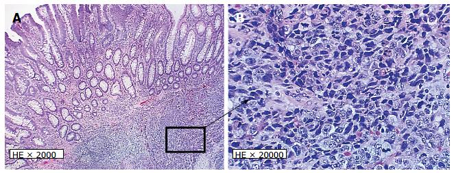
Figure 2 Hematoxylin and eosin staining of the tumor sections at × 2000 (A) and × 20000 (B).
A: Shows the tubular adenoma component juxtaposed to the underlying high-grade large-cell neuroendocrinal carcinoma; B: Higher magnification showing medium to large-sized tumor cells with vesicular nuclei, prominent nucleoli consistent with large-cell neuroendocrine carcinoma.
- Citation: Soliman ML, Tiwari A, Zhao Q. Coexisting tubular adenoma with a neuroendocrine carcinoma of colon allowing early surgical intervention and implicating a shared stem cell origin. World J Gastroenterol 2017; 23(6): 1106-1112
- URL: https://www.wjgnet.com/1007-9327/full/v23/i6/1106.htm
- DOI: https://dx.doi.org/10.3748/wjg.v23.i6.1106


