Copyright
©The Author(s) 2017.
World J Gastroenterol. Feb 14, 2017; 23(6): 1018-1029
Published online Feb 14, 2017. doi: 10.3748/wjg.v23.i6.1018
Published online Feb 14, 2017. doi: 10.3748/wjg.v23.i6.1018
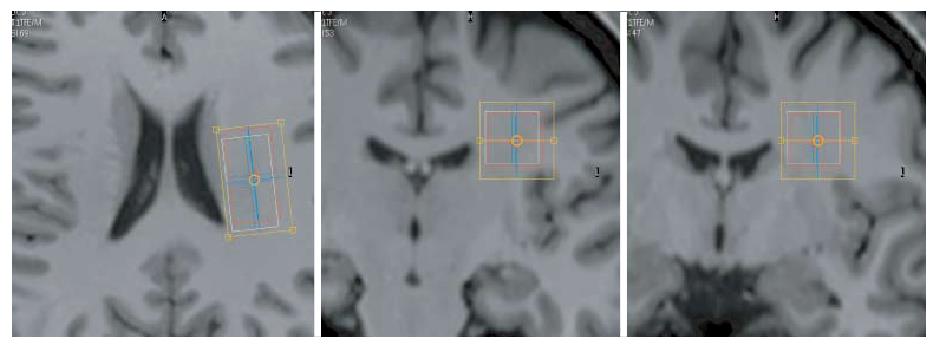
Figure 2 Planning of the 1H-magnetic resonance spectroscopy volume of interest in the left centrum semi-ovale.
Seen on axial (left) and on the coronal (right) T1-weighted image slices. The effective volume of interest set at the tNAA frequency is shown (red rectangle) together with the shimming volume (yellow rectangle).
- Citation: van Erp S, Ercan E, Breedveld P, Brakenhoff L, Ghariq E, Schmid S, Osch MV, van Buchem M, Emmer B, van der Grond J, Wolterbeek R, Hommes D, Fidder H, van der Wee N, Huizinga T, van der Heijde D, Middelkoop H, Ronen I, van der Meulen-de Jong A. Cerebral magnetic resonance imaging in quiescent Crohn’s disease patients with fatigue. World J Gastroenterol 2017; 23(6): 1018-1029
- URL: https://www.wjgnet.com/1007-9327/full/v23/i6/1018.htm
- DOI: https://dx.doi.org/10.3748/wjg.v23.i6.1018


