Copyright
©The Author(s) 2017.
World J Gastroenterol. Feb 7, 2017; 23(5): 919-925
Published online Feb 7, 2017. doi: 10.3748/wjg.v23.i5.919
Published online Feb 7, 2017. doi: 10.3748/wjg.v23.i5.919
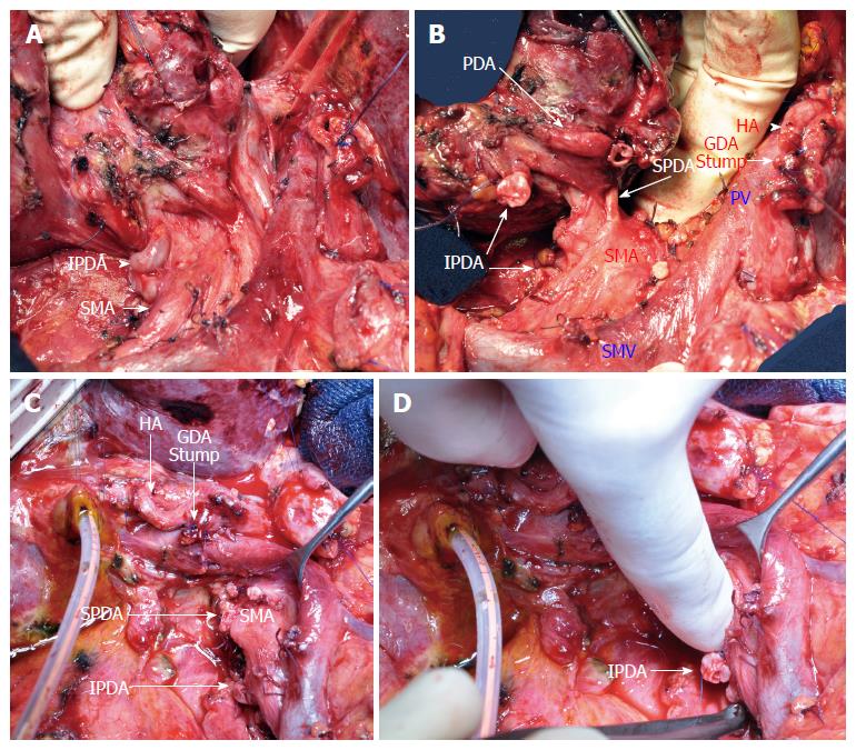
Figure 4 Pancreaticoduodenectomy.
Operative views (A, B) after mobilization of the specimen showing a very large inferior pancreaticoduodenal artery (IPDA) and the pancreaticoduodenal arcade. After resection (C, D), the stumps of the pancreaticoduodenal arteries (superior pancreaticoduodenal artery and IPDA) are shown. SMA: Superior mesenteric artery; HA: Hepatic artery; GDA: Gastroduodenal artery; SMV: Superior mesenteric vein; PV: Portal vein.
- Citation: Guilbaud T, Ewald J, Turrini O, Delpero JR. Pancreaticoduodenectomy: Secondary stenting of the celiac trunk after inefficient median arcuate ligament release and reoperation as an alternative to simultaneous hepatic artery reconstruction. World J Gastroenterol 2017; 23(5): 919-925
- URL: https://www.wjgnet.com/1007-9327/full/v23/i5/919.htm
- DOI: https://dx.doi.org/10.3748/wjg.v23.i5.919


