Copyright
©The Author(s) 2017.
World J Gastroenterol. Feb 7, 2017; 23(5): 906-918
Published online Feb 7, 2017. doi: 10.3748/wjg.v23.i5.906
Published online Feb 7, 2017. doi: 10.3748/wjg.v23.i5.906
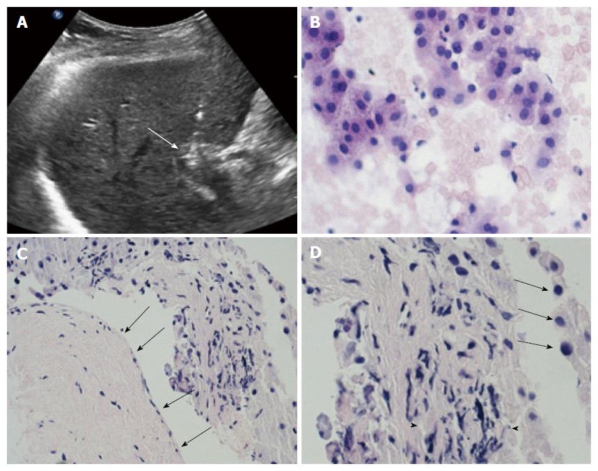
Figure 4 All patients underwent pre- and post-treatment biopsy of the portal vein tumor thrombus.
A: ultrasound scan demonstrate the correct positioning of the needle tip (arrow) in the thrombus; B: Intraoperative pre-treatment biopsy of the thrombus was adequate and showed viable cells from hepatocellular carcinoma in 5/6 cases; C: High magnification of biopsy specimen showed severe involutive changes of tumor cells with cellular apoptosis (arrows) and areas of necrosis (arrowheads) in all six cases. Low magnification in the same specimen, beside the altered tumor thrombus, showed absence of damage from the procedure to portal vein wall (arrows); D: Portal endothelium shows normal appearance with regular wall layers.
- Citation: Tarantino L, Busto G, Nasto A, Fristachi R, Cacace L, Talamo M, Accardo C, Bortone S, Gallo P, Tarantino P, Nasto RA, Di Minno MND, Ambrosino P. Percutaneous electrochemotherapy in the treatment of portal vein tumor thrombosis at hepatic hilum in patients with hepatocellular carcinoma in cirrhosis: A feasibility study. World J Gastroenterol 2017; 23(5): 906-918
- URL: https://www.wjgnet.com/1007-9327/full/v23/i5/906.htm
- DOI: https://dx.doi.org/10.3748/wjg.v23.i5.906


